
p70 S6 Kinase 2 Rabbit mAb, Unconjugated, Monoclonal
Catalog Number:
ABB-A9100
- Images (9)

| Article Name: | p70 S6 Kinase 2 Rabbit mAb, Unconjugated, Monoclonal |
| Biozol Catalog Number: | ABB-A9100 |
| Supplier Catalog Number: | A9100 |
| Alternative Catalog Number: | ABB-A9100-20UL,ABB-A9100-100UL |
| Manufacturer: | ABclonal |
| Host: | Rabbit |
| Category: | Antikörper |
| Application: | ELISA, IF, IHC-P, WB |
| Species Reactivity: | Human |
| Immunogen: | Recombinant protein (or fragment).This information is considered to be commercially sensitive. |
| Conjugation: | Unconjugated |
| Alternative Names: | KLS, SRK, S6K2, S6KB, STK14B, S6KI(2), S6Kbeta, p70S6Kb, P70-beta, S6K-beta2, P70-beta-1, P70-beta-2, p70(S6K)-beta, p70 S6 Kinase 2 |
| This gene encodes a member of the RSK (ribosomal S6 kinase) family of serine/threonine kinases. This kinase contains a kinase catalytic domain and phosphorylates the S6 ribosomal protein and eukaryotic translation initiation factor 4B (eIF4B). Phosphorylation of S6 leads to an increase in protein synthesis and cell proliferation. |
| Application Dilute: | WB,1:500 - 1:2000|IHC-P,1:50 - 1:200|IF/ICC,1:50 - 1:200|ELISA,Recommended starting concentration is 1 µg/mL. Please optimize the concentration based on your specific assay requirements. |
| Application Notes: | Cross-Reactivity: Human,Mouse,Rat. ResearchArea: Epigenetics Nuclear Signaling,Translation Control,Regulation of eIF4 and p70 S6 Kinase,Signal Transduction,Kinase,Serine threonine kinases,PI3K-Akt Signaling Pathway,mTOR Signaling Pathway,ErbB-HER Signaling Pathway,Cell Biology Developmental Biology,Apoptosis,Mitochondrial Control of Apoptosis,Inhibition of Apoptosis,TGF-b-Smad Signaling Pathway,Endocrine Metabolism,AMPK Signaling Pathway,Insulin Receptor Signaling Pathway,Endocrine and metabolic diseases,Diabetes,Obesity,Immunology Inflammation,B Cell Receptor Signaling Pathway,Cardiovascular,Angiogenesis. Shipping: Ice Bag |
|
|
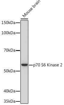
Western blot analysis of lysates from Mouse brain, using p70 S6 Kinase 2 Rabbit mAb (A9100) at 1:1000 dilution incubated overnight at 4°C. Secondary antibody: HRP-conjugated Goat anti-Rabbit IgG (H+L) (AS014) at 1:10000 dilution. Lysates/proteins: 25 µg per lane. Blocking buffer: 3% nonfat dry milk in TBST. Detection: ECL Basic Kit (RM00020). Exposure time: 3 s. |
|
|
Western blot analysis of lysates from A549 cells using p70 S6 Kinase 2 Rabbit mAb (A9100) at 1:1000 dilution incubated overnight at 4°C. Secondary antibody: HRP-conjugated Goat anti-Rabbit IgG (H+L) (AS014) at 1:10000 dilution. Lysates/proteins: 25 µg per lane. Blocking buffer: 3% nonfat dry milk in TBST. Detection: ECL Basic Kit (RM00020). Exposure time: 30 s. |
|
|
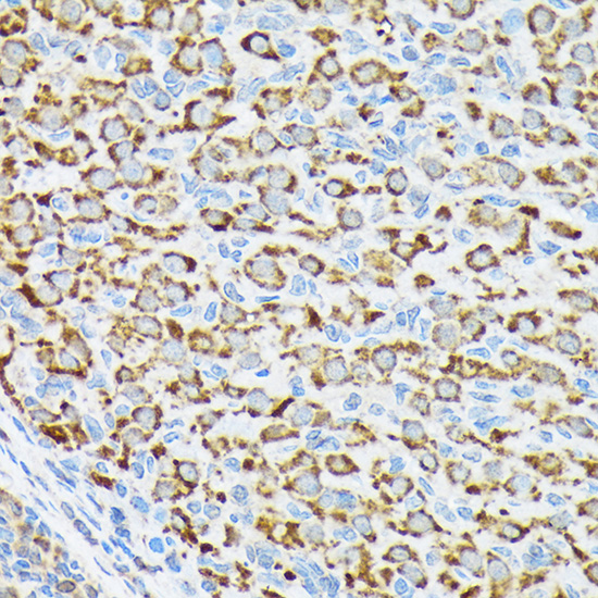
Immunohistochemistry analysis of paraffin-embeddedHuman colon carcinoma tissue usingp70 S6 Kinase 2 Rabbit mAb(A9100) at a dilution of 1:200 (40x lens).High pressure antigen retrieval was performed with 0.01 M citrate buffer (pH 6.0) prior to IHC staining. |
|
|
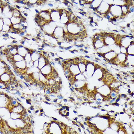
Immunohistochemistry analysis of paraffin-embeddedRat colon tissue usingp70 S6 Kinase 2 Rabbit mAb(A9100) at a dilution of 1:200 (40x lens).High pressure antigen retrieval was performed with 0.01 M citrate buffer (pH 6.0) prior to IHC staining. |
|
|
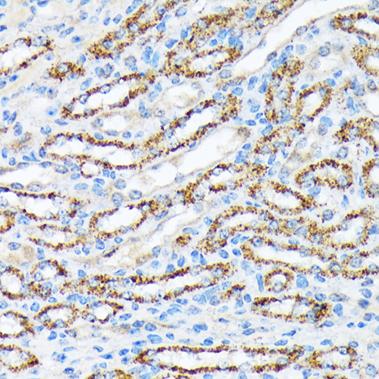
Immunohistochemistry analysis of paraffin-embeddedMouse kidney tissue usingp70 S6 Kinase 2 Rabbit mAb(A9100) at a dilution of 1:200 (40x lens).High pressure antigen retrieval was performed with 0.01 M citrate buffer (pH 6.0) prior to IHC staining. |
|
|
Immunohistochemistry analysis of paraffin-embeddedHuman cervix cancer tissue usingp70 S6 Kinase 2 Rabbit mAb(A9100) at a dilution of 1:200 (40x lens).High pressure antigen retrieval was performed with 0.01 M citrate buffer (pH 6.0) prior to IHC staining. |
|
|
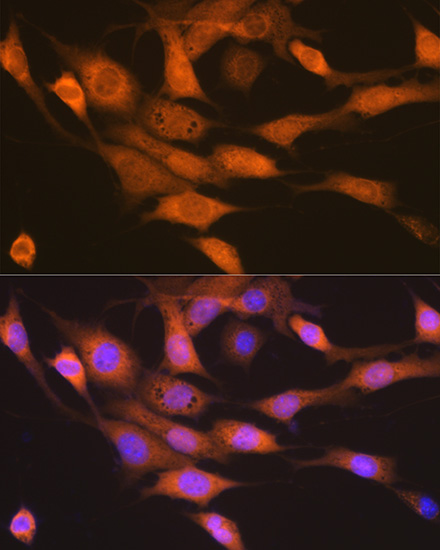
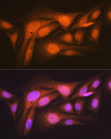
Confocal imaging of C2C12 cells usingp70 S6 Kinase 2 Rabbit mAb (A9100, dilution 1:100) followed by a further incubation with Cy3 Goat Anti-Rabbit IgG (H+L) (AS007, dilution 1:500) (Red). The cells were counterstained with alpha-Tubulin Mouse mAb (AC012, dilution 1:400) followed by incubation with ABflo 488-conjugated Goat Anti-Mouse IgG (H+L) Ab (AS076, dilution 1:500) (Green). DAPI was used for nuclear staining (Blue). Objective: 100x. |
|
|
Confocal imaging of NIH/3T3 cells usingp70 S6 Kinase 2 Rabbit mAb (A9100, dilution 1:100) followed by a further incubation with Cy3 Goat Anti-Rabbit IgG (H+L) (AS007, dilution 1:500) (Red). The cells were counterstained with alpha-Tubulin Mouse mAb (AC012, dilution 1:400) followed by incubation with ABflo 488-conjugated Goat Anti-Mouse IgG (H+L) Ab (AS076, dilution 1:500) (Green). DAPI was used for nuclear staining (Blue). Objective: 100x. |
|
|
Confocal imaging of U-2 OS cells usingp70 S6 Kinase 2 Rabbit mAb (A9100,dilution 1:100) followed by a further incubation with Cy3 Goat Anti-Rabbit IgG (H+L) (AS007, dilution 1:500) (Red). The cells were counterstained with alpha-Tubulin Mouse mAb (AC012, dilution 1:400) followed by incubation with ABflo 488-conjugated Goat Anti-Mouse IgG (H+L) Ab (AS076, dilution 1:500) (Green). DAPI was used for nuclear staining (Blue). Objective: 100x. |
Product Guarantee and Expert Support
