
RanGAP1 Rabbit mAb, Unconjugated, Monoclonal
Catalog Number:
ABB-A8758
- Images (9)

| Article Name: | RanGAP1 Rabbit mAb, Unconjugated, Monoclonal |
| Biozol Catalog Number: | ABB-A8758 |
| Supplier Catalog Number: | A8758 |
| Alternative Catalog Number: | ABB-A8758-20UL,ABB-A8758-100UL |
| Manufacturer: | ABclonal |
| Host: | Rabbit |
| Category: | Antikörper |
| Application: | ELISA, IF, IHC-P, WB |
| Species Reactivity: | Human |
| Immunogen: | Synthetic peptide. This information is considered to be commercially sensitive. |
| Conjugation: | Unconjugated |
| Alternative Names: | SD, Fug1, RANGAP, RanGAP1 |
| This gene encodes a protein that associates with the nuclear pore complex and participates in the regulation of nuclear transport. The encoded protein interacts with Ras-related nuclear protein 1 (RAN) and regulates guanosine triphosphate (GTP)-binding and exchange. Alternative splicing results in multiple transcript variants. |
| Application Dilute: | WB,1:500 - 1:2000|IHC-P,1:50 - 1:200|IF/ICC,1:50 - 1:200|ELISA,Recommended starting concentration is 1 µg/mL. Please optimize the concentration based on your specific assay requirements. |
| Application Notes: | Cross-Reactivity: Human,Mouse,Rat. ResearchArea: Epigenetics Nuclear Signaling,Nuclear Receptor Signaling,Cell Biology Developmental Biology,Cell Cycle. Shipping: Ice Bag |
|
|
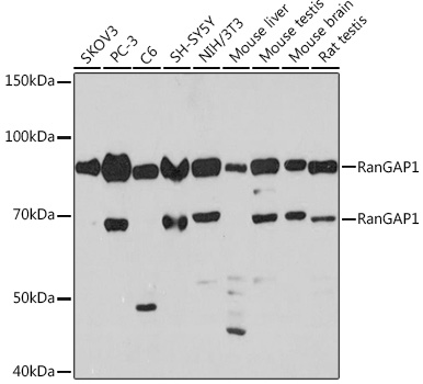
Western blot analysis of various lysates using RanGAP1 Rabbit mAb (A8758) at 1:1000 dilution. Secondary antibody: HRP-conjugated Goat anti-Rabbit IgG (H+L) (AS014) at 1:10000 dilution. Lysates/proteins: 25µg per lane. Blocking buffer: 3% nonfat dry milk in TBST. Detection: ECL Basic Kit (RM00020). Exposure time: 3min. |
|
|
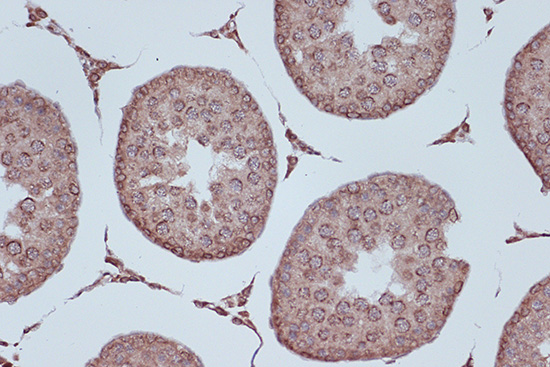
Immunohistochemistry analysis of paraffin-embeddedRat testis tissue usingRanGAP1 Rabbit mAb(A8758) at a dilution of 1:200 (40x lens).High pressure antigen retrieval was performed with 0.01 M citrate buffer (pH 6.0) prior to IHC staining. |
|
|
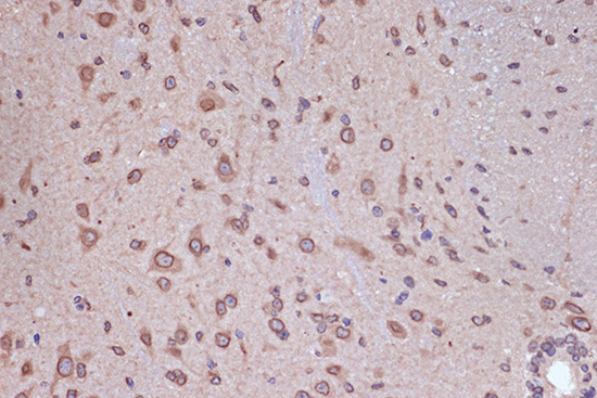
Immunohistochemistry analysis of paraffin-embeddedMouse brain tissue usingRanGAP1 Rabbit mAb(A8758) at a dilution of 1:200 (40x lens).High pressure antigen retrieval was performed with 0.01 M citrate buffer (pH 6.0) prior to IHC staining. |
|
|
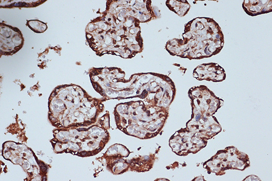
Immunohistochemistry analysis of paraffin-embeddedHuman colon carcinoma tissue usingRanGAP1 Rabbit mAb(A8758) at a dilution of 1:200 (40x lens).High pressure antigen retrieval was performed with 0.01 M citrate buffer (pH 6.0) prior to IHC staining. |
|
|
Immunohistochemistry analysis of paraffin-embeddedRat liver tissue usingRanGAP1 Rabbit mAb(A8758) at a dilution of 1:200 (40x lens).High pressure antigen retrieval was performed with 0.01 M citrate buffer (pH 6.0) prior to IHC staining. |
|
|
Immunohistochemistry analysis of paraffin-embeddedMouse kidney tissue usingRanGAP1 Rabbit mAb(A8758) at a dilution of 1:200 (40x lens).High pressure antigen retrieval was performed with 0.01 M citrate buffer (pH 6.0) prior to IHC staining. |
|
|
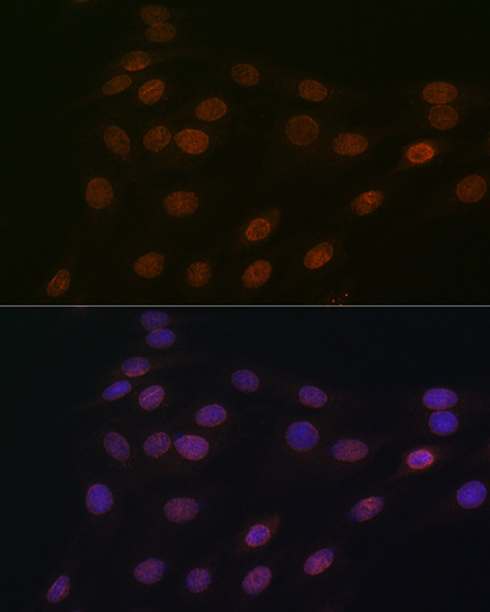
Immunofluorescence analysis of C6 cells using RanGAP1 Rabbit mAb (A8758) at dilution of 1:100 (40x lens). Secondary antibody: Cy3-conjugated Goat anti-Rabbit IgG (H+L) (AS007) at 1:500 dilution. Blue: DAPI for nuclear staining. |
|
|
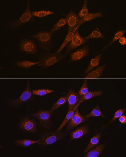
Immunofluorescence analysis of NIH-3T3 cells using RanGAP1 Rabbit mAb (A8758) at dilution of 1:100 (40x lens). Secondary antibody: Cy3-conjugated Goat anti-Rabbit IgG (H+L) (AS007) at 1:500 dilution. Blue: DAPI for nuclear staining. |
|
|
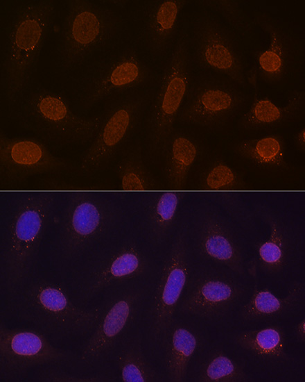
Immunofluorescence analysis of U-2 OS cells using RanGAP1 Rabbit mAb (A8758) at dilution of 1:100 (40x lens). Secondary antibody: Cy3-conjugated Goat anti-Rabbit IgG (H+L) (AS007) at 1:500 dilution. Blue: DAPI for nuclear staining. |
Product Guarantee and Expert Support
