
IKZF3 Rabbit mAb, Unconjugated, Monoclonal
Catalog Number:
ABB-A8614
- Images (8)
| Article Name: | IKZF3 Rabbit mAb, Unconjugated, Monoclonal |
| Biozol Catalog Number: | ABB-A8614 |
| Supplier Catalog Number: | A8614 |
| Alternative Catalog Number: | ABB-A8614-100UL,ABB-A8614-20UL |
| Manufacturer: | ABclonal |
| Host: | Rabbit |
| Category: | Antikörper |
| Application: | ELISA, IF, IHC-P, IP, WB |
| Species Reactivity: | Human |
| Immunogen: | Recombinant protein (or fragment).This information is considered to be commercially sensitive. |
| Conjugation: | Unconjugated |
| Alternative Names: | AIO, IMD84, AIOLOS, ZNFN1A3, IKZF3 |
| This gene encodes a member of the Ikaros family of zinc-finger proteins. Three members of this protein family (Ikaros, Aiolos and Helios) are hematopoietic-specific transcription factors involved in the regulation of lymphocyte development. This gene product is a transcription factor that is important in the regulation of B lymphocyte proliferation and differentiation. Both Ikaros and Aiolos can participate in chromatin remodeling. Regulation of gene expression in B lymphocytes by Aiolos is complex as it appears to require the sequential formation of Ikaros homodimers, Ikaros/Aiolos heterodimers, and Aiolos homodimers. Several alternative transcripts encoding different isoforms have been described, as well as some non-protein coding variants. |
| Application Dilute: | WB,1:500 - 1:1000|IP,0.5µg-4µg antibody for 200µg-400µg extracts of whole cells|IF-P,1:50 - 1:200|IHC-P,1:200 - 1:800|ELISA,Recommended starting concentration is 1 µg/mL. Please optimize the concentration based on your specific assay requirements. |
| Application Notes: | Cross-Reactivity: Human,Mouse,Rat. ResearchArea: Epigenetics Nuclear Signaling,Transcription Factors. Shipping: Ice Bag |
|
|
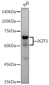
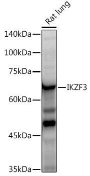
Western blot analysis of various lysates using IKZF3 Rabbit mAb (A8614) at 1:500 dilution. Secondary antibody: HRP-conjugated Goat anti-Rabbit IgG (H+L) (AS014) at 1:10000 dilution. Lysates/proteins: 25µg per lane. Blocking buffer: 3% nonfat dry milk in TBST. Detection: ECL Basic Kit (RM00020). Exposure time: 60s. |
|
|
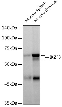
Western blot analysis of various lysates using IKZF3 Rabbit mAb (A8614) at 1:1000 dilutionincubated at room temperature for 1.5 hours. Secondary antibody: HRP-conjugated Goat anti-Rabbit IgG (H+L) (AS014) at 1:10000 dilution. Lysates/proteins: 25 µg per lane. Blocking buffer: 3% nonfat dry milk in TBST. Detection: ECL Basic Kit (RM00020). Negative control (NC): U-937. Exposure time: 45 s. |
|
|
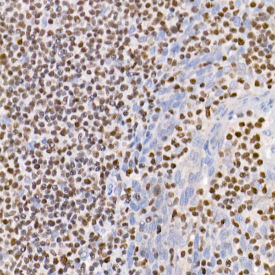
Immunohistochemistry analysis of paraffin-embedded Human spleen tissue using IKZF3 Rabbit mAb (A8614) at a dilution of 1:200 (40x lens). High pressure antigen retrieval performed with 0.01M Citrate buffer (pH 6.0) prior to IHC staining. |
|
|
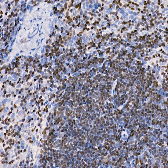
Immunohistochemistry analysis of paraffin-embedded Mouse spleen tissue using IKZF3 Rabbit mAb (A8614) at a dilution of 1:200 (40x lens). High pressure antigen retrieval performed with 0.01M Citrate buffer (pH 6.0) prior to IHC staining. |
|
|
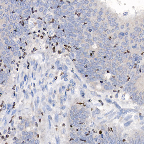
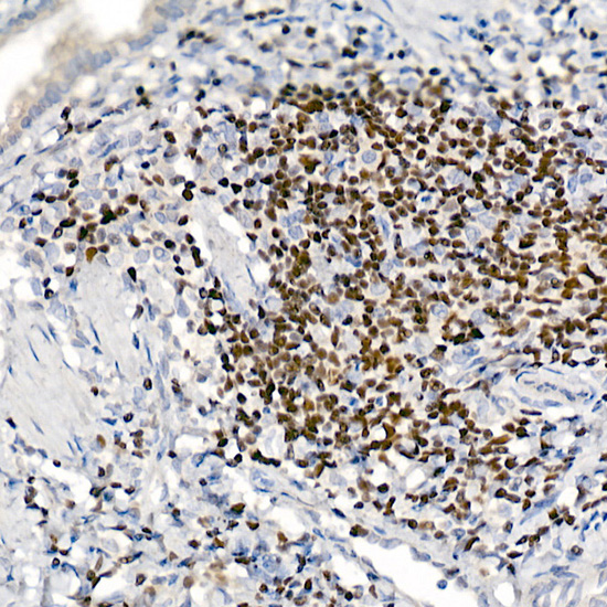
Immunohistochemistry analysis of paraffin-embedded Rat spleen tissue using IKZF3 Rabbit mAb (A8614) at a dilution of 1:200 (40x lens). High pressure antigen retrieval performed with 0.01M Citrate buffer (pH 6.0) prior to IHC staining. |
|
|
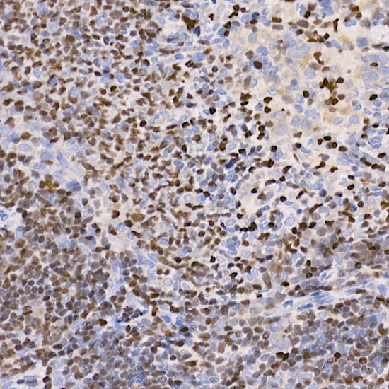
Immunofluorescence analysis of paraffin-embedded rat spleen using IKZF3 Rabbit mAb (A8614) at dilution of 1:100 (40x lens). Secondary antibody: Cy3-conjugated Goat anti-Rabbit IgG (H+L) (AS007) at 1:500 dilution. Blue: DAPI for nuclear staining. |
|
|
Immunoprecipitation analysis of 300 µg extracts of Raji cells using 3 µg IKZF3 antibody (A8614). Western blot was performed from the immunoprecipitate using IKZF3 antibody (A8614) at a dilution of 1:500. |
|
|
Immunoprecipitation analysis of 300 µg extracts of Raji cells using 3 µg IKZF3 antibody (A8614). Western blot was performed from the immunoprecipitate using IKZF3 antibody (A8614) at a dilution of 1:500. |
Product Guarantee and Expert Support
