
[KD Validated] TFEB Rabbit pAb, Unconjugated, Polyclonal
Catalog Number:
ABB-A7311
- Images (8)


| Article Name: | [KD Validated] TFEB Rabbit pAb, Unconjugated, Polyclonal |
| Biozol Catalog Number: | ABB-A7311 |
| Supplier Catalog Number: | A7311 |
| Alternative Catalog Number: | ABB-A7311-100UL,ABB-A7311-20UL,ABB-A7311-500UL,ABB-A7311-1000UL |
| Manufacturer: | ABclonal |
| Host: | Rabbit |
| Category: | Antikörper |
| Application: | ELISA, IF, IHC-P, WB |
| Species Reactivity: | Human |
| Immunogen: | Recombinant protein (or fragment).This information is considered to be commercially sensitive. |
| Conjugation: | Unconjugated |
| Alternative Names: | TCFEB, BHLHE35, ALPHATFEB, TFEB |
| Enables DNA-binding transcription factor activity, enzyme binding activity, and transcription cis-regulatory region binding activity. Involved in several processes, including cellular response to amino acid starvation, lysosome localization, and positive regulation of autophagy. Located in cytosol, lysosomal membrane, and nucleoplasm. |
| Application Dilute: | WB,1:500 - 1:5000|IF/ICC,1:50 - 1:200|IHC-P,1:50 - 1:200|ELISA,Recommended starting concentration is 1 µg/mL. Please optimize the concentration based on your specific assay requirements. |
| Application Notes: | Cross-Reactivity: Human,Mouse,Rat. ResearchArea: Epigenetics Nuclear Signaling,Transcription Factors. Shipping: Ice Bag |
|
|
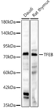
Western blot analysis of lysates from wild type (WT) and TFEB knockdown (KD) HeLa cells using [KD Validated] TFEB Rabbit pAb (A7311) at 1:1000 dilution incubated overnight at 4°C. Secondary antibody: HRP-conjugated Goat anti-Rabbit IgG (H+L) (AS014) at 1:10000 dilution. Lysates/proteins: 25 µg per lane. Blocking buffer: 3% nonfat dry milk in TBST. Detection: ECL Basic Kit (RM00020). Exposure time: 10s. |
|
|
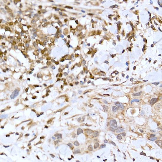
Immunohistochemistry analysis of paraffin-embeddedHuman colon tissue using [KD Validated]TFEB Rabbit pAb(A7311) at a dilution of 1:100 (40x lens).High pressure antigen retrieval was performed with 0.01 M citrate buffer (pH 6.0) prior to IHC staining. |
|
|
Western blot analysis of various lysates, using [KD Validated] TFEB Rabbit pAb (A7311) at 1:1000 dilution. Secondary antibody: HRP-conjugated Goat anti-Rabbit IgG (H+L) (AS014) at 1:10000 dilution. Lysates/proteins: 25µg per lane. Blocking buffer: 3% nonfat dry milk in TBST. Detection: ECL Basic Kit (RM00020). Exposure time: 180s. |
|
|
Immunohistochemistry analysis of paraffin-embeddedMouse lung tissue using [KD Validated]TFEB Rabbit pAb(A7311) at a dilution of 1:100 (40x lens).High pressure antigen retrieval was performed with 0.01 M citrate buffer (pH 6.0) prior to IHC staining. |
|
|
Immunohistochemistry analysis of paraffin-embeddedRat testis tissue using [KD Validated]TFEB Rabbit pAb(A7311) at a dilution of 1:100 (40x lens).High pressure antigen retrieval was performed with 0.01 M citrate buffer (pH 6.0) prior to IHC staining. |
|
|
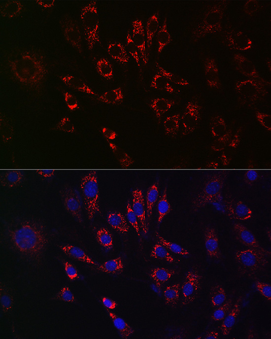
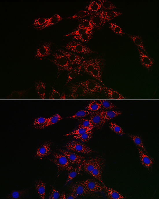
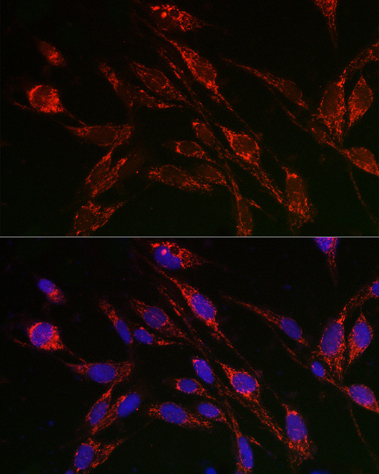
Immunofluorescence analysis of NIH/3T3 cells using [KD Validated] TFEB Rabbit pAb (A7311) at dilution of 1:50 (40x lens). Secondary antibody: Cy3-conjugated Goat anti-Rabbit IgG (H+L) (AS007) at 1:500 dilution. Blue: DAPI for nuclear staining. |
|
|
Immunofluorescence analysis of PC-12 cells using [KD Validated] TFEB Rabbit pAb (A7311) at dilution of 1:50 (40x lens). Secondary antibody: Cy3-conjugated Goat anti-Rabbit IgG (H+L) (AS007) at 1:500 dilution. Blue: DAPI for nuclear staining. |
|
|
Immunofluorescence analysis of U2OS cells using [KD Validated] TFEB Rabbit pAb (A7311) at dilution of 1:50 (40x lens). Secondary antibody: Cy3-conjugated Goat anti-Rabbit IgG (H+L) (AS007) at 1:500 dilution. Blue: DAPI for nuclear staining. |
Product Guarantee and Expert Support
