
FABP5 Rabbit pAb, Unconjugated, Polyclonal
Catalog Number:
ABB-A6373
- Images (9)

| Article Name: | FABP5 Rabbit pAb, Unconjugated, Polyclonal |
| Biozol Catalog Number: | ABB-A6373 |
| Supplier Catalog Number: | A6373 |
| Alternative Catalog Number: | ABB-A6373-20UL,ABB-A6373-100UL,ABB-A6373-500UL,ABB-A6373-1000UL |
| Manufacturer: | ABclonal |
| Host: | Rabbit |
| Category: | Antikörper |
| Application: | ELISA, IF, IHC-P, WB |
| Species Reactivity: | Human |
| Immunogen: | Recombinant protein (or fragment).This information is considered to be commercially sensitive. |
| Conjugation: | Unconjugated |
| Alternative Names: | EFABP, KFABP, E-FABP, PAFABP, PA-FABP, FABP5 |
| This gene encodes the fatty acid binding protein found in epidermal cells, and was first identified as being upregulated in psoriasis tissue. Fatty acid binding proteins are a family of small, highly conserved, cytoplasmic proteins that bind long-chain fatty acids and other hydrophobic ligands. FABPs may play roles in fatty acid uptake, transport, and metabolism. Polymorphisms in this gene are associated with type 2 diabetes. The human genome contains many pseudogenes similar to this locus. |
| Application Dilute: | WB,1:500 - 1:1000|IHC-P,1:50 - 1:200|IF/ICC,1:50 - 1:200|ELISA,Recommended starting concentration is 1 µg/mL. Please optimize the concentration based on your specific assay requirements. |
| Application Notes: | Cross-Reactivity: Human,Mouse,Rat. ResearchArea: Cancer,Signal Transduction,Endocrine Metabolism,Lipid Metabolism,Cardiovascular,Lipids,Fatty Acids. Shipping: Ice Bag |
|
|
Immunohistochemistry analysis of paraffin-embedded Human kidney tissue using FABP5 Rabbit pAb (A6373) at a dilution of 1:300 (40x lens). High pressure antigen retrieval performed with 0.01M Tris-EDTA Buffer (pH 9.0) prior to IHC staining. |
|
|
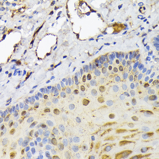
Immunohistochemistry analysis of paraffin-embedded Human cervix cancer tissue using FABP5 Rabbit pAb (A6373) at a dilution of 1:300 (40x lens). High pressure antigen retrieval performed with 0.01M Tris-EDTA Buffer (pH 9.0) prior to IHC staining. |
|
|
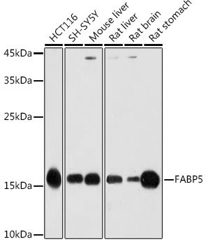
Western blot analysis of various lysates using FABP5 Rabbit pAb (A6373) at 1:1000 dilution. Secondary antibody: HRP-conjugated Goat anti-Rabbit IgG (H+L) (AS014) at 1:10000 dilution. Lysates/proteins: 25µg per lane. Blocking buffer: 3% nonfat dry milk in TBST. Detection: ECL Basic Kit (RM00020). Exposure time: 30s. |
|
|
Immunohistochemistry analysis of paraffin-embedded Human lung tissue using FABP5 Rabbit pAb (A6373) at a dilution of 1:300 (40x lens). High pressure antigen retrieval performed with 0.01M Tris-EDTA Buffer (pH 9.0) prior to IHC staining. |
|
|
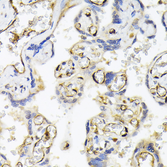
Immunohistochemistry analysis of paraffin-embedded Human placenta tissue using FABP5 Rabbit pAb (A6373) at a dilution of 1:300 (40x lens). High pressure antigen retrieval performed with 0.01M Tris-EDTA Buffer (pH 9.0) prior to IHC staining. |
|
|
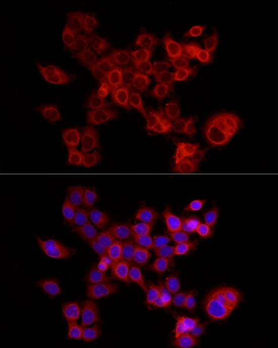
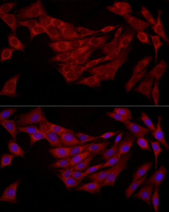
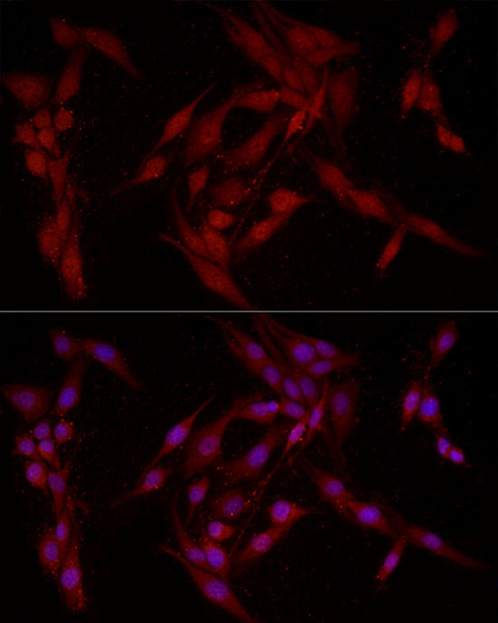
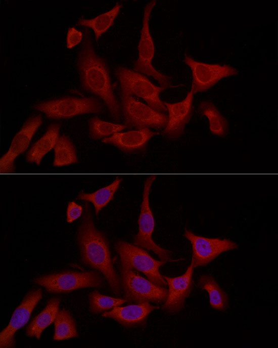
Immunofluorescence analysis of HCT116 cells using FABP5 Rabbit pAb (A6373) at dilution of 1:200 (40x lens). Secondary antibody: Cy3-conjugated Goat anti-Rabbit IgG (H+L) (AS007) at 1:500 dilution. Blue: DAPI for nuclear staining. |
|
|
Immunofluorescence analysis of NIH/3T3 cells using FABP5 Rabbit pAb (A6373) at dilution of 1:200 (40x lens). Secondary antibody: Cy3-conjugated Goat anti-Rabbit IgG (H+L) (AS007) at 1:500 dilution. Blue: DAPI for nuclear staining. |
|
|
Immunofluorescence analysis of PC-12 cells using FABP5 Rabbit pAb (A6373) at dilution of 1:200 (40x lens). Secondary antibody: Cy3-conjugated Goat anti-Rabbit IgG (H+L) (AS007) at 1:500 dilution. Blue: DAPI for nuclear staining. |
|
|
Immunofluorescence analysis of U2OS cells using FABP5 Rabbit pAb (A6373) at dilution of 1:200 (40x lens). Secondary antibody: Cy3-conjugated Goat anti-Rabbit IgG (H+L) (AS007) at 1:500 dilution. Blue: DAPI for nuclear staining. |
Product Guarantee and Expert Support
