
RPL17 Rabbit pAb, Unconjugated, Polyclonal
Catalog Number:
ABB-A5934
- Images (8)


| Article Name: | RPL17 Rabbit pAb, Unconjugated, Polyclonal |
| Biozol Catalog Number: | ABB-A5934 |
| Supplier Catalog Number: | A5934 |
| Alternative Catalog Number: | ABB-A5934-20UL,ABB-A5934-100UL,ABB-A5934-500UL,ABB-A5934-1000UL |
| Manufacturer: | ABclonal |
| Host: | Rabbit |
| Category: | Antikörper |
| Application: | ELISA, IF, IHC-P, WB |
| Species Reactivity: | Human |
| Immunogen: | Recombinant protein (or fragment).This information is considered to be commercially sensitive. |
| Conjugation: | Unconjugated |
| Alternative Names: | L17, PD-1, uL22, RPL23, RPL17 |
| Ribosomes, the organelles that catalyze protein synthesis, consist of a small 40S subunit and a large 60S subunit. Together these subunits are composed of 4 RNA species and approximately 80 structurally distinct proteins. This gene encodes a ribosomal protein that is a component of the 60S subunit. The protein belongs to the L22P family of ribosomal proteins. It is located in the cytoplasm. This gene has been referred to as rpL23 because the encoded protein shares amino acid identity with ribosomal protein L23 from Halobacterium marismortui, however, its official symbol is RPL17. As is typical for genes encoding ribosomal proteins, there are multiple processed pseudogenes of this gene dispersed through the genome. Alternative splicing results in multiple transcript variants. Read-through transcription also exists between this gene and the neighboring downstream C18orf32 (chromosome 18 open reading frame 32) gene. |
| Clonality: | Polyclonal |
| Molecular Weight: | 21kDa |
| NCBI: | 6139 |
| UniProt: | P18621 |
| Purity: | Affinity purification |
| Sequence: | MVRYSLDPENPTKSCKSRGSNLRVHFKNTRETAQAIKGMHIRKATKYLKDVTLQKQCVPFRRYNGGVGRCAQAKQWGWTQGRWPKKSAEFLLHMLKNAESNAELKGLDVDSLVIEHIQVNKAPKMRRRTYRAHGRINPYMSSPCHIEMILTEKEQIVPKPEEEVAQKKKISQKKLKKQKLMARE |
| Target: | RPL17 |
| Antibody Type: | Primary Antibody |
| Application Dilute: | WB,1:200 - 1:2000|IHC-P,1:50 - 1:200|IF/ICC,1:50 - 1:200|ELISA,Recommended starting concentration is 1 µg/mL. Please optimize the concentration based on your specific assay requirements. |
| Application Notes: | Cross-Reactivity: Human,Mouse,Rat. ResearchArea: Epigenetics Nuclear Signaling,RNA Binding. Shipping: Ice Bag |
|
|
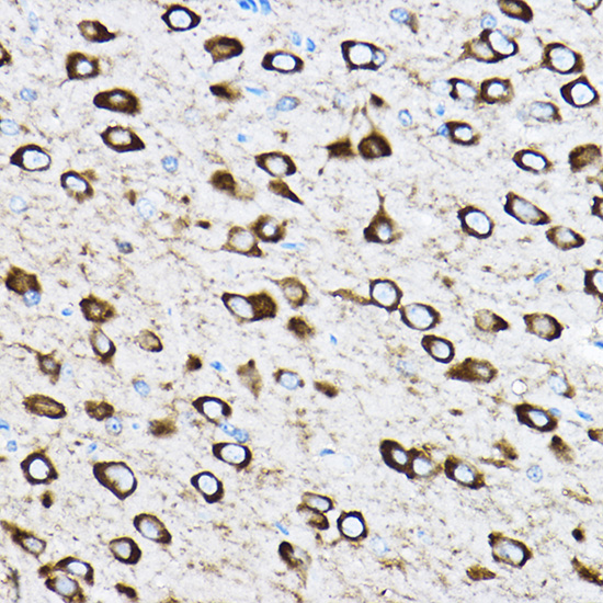
Immunohistochemistry analysis of paraffin-embedded Rat brain using RPL17 Rabbit pAb (A5934) at dilution of 1:100 (40x lens). High pressure antigen retrieval performed with 0.01M Citrate buffer (pH 6.0) prior to IHC staining. |
|
|
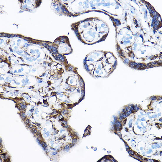
Immunohistochemistry analysis of paraffin-embedded Human placenta using RPL17 Rabbit pAb (A5934) at dilution of 1:100 (40x lens). High pressure antigen retrieval performed with 0.01M Citrate buffer (pH 6.0) prior to IHC staining. |
|
|
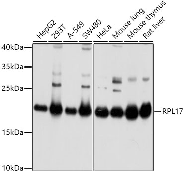
Western blot analysis of various lysates using RPL17 Rabbit pAb (A5934) at 1:1000 dilution. Secondary antibody: HRP-conjugated Goat anti-Rabbit IgG (H+L) (AS014) at 1:10000 dilution. Lysates/proteins: 25µg per lane. Blocking buffer: 3% nonfat dry milk in TBST. Detection: ECL Basic Kit (RM00020). Exposure time: 5s. |
|
|
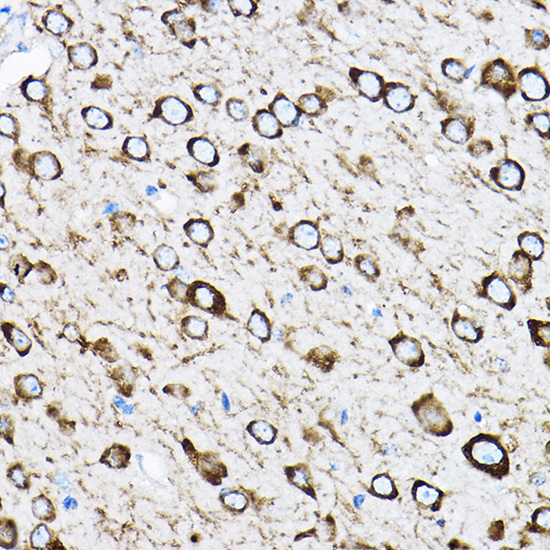
Immunohistochemistry analysis of paraffin-embedded Mouse brain using RPL17 Rabbit pAb (A5934) at dilution of 1:100 (40x lens). High pressure antigen retrieval performed with 0.01M Citrate buffer (pH 6.0) prior to IHC staining. |
|
|
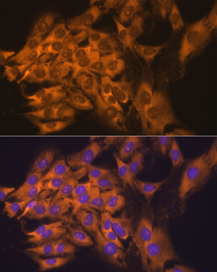
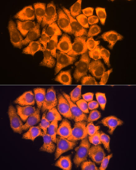
Immunofluorescence analysis of C6 cells using RPL17 Rabbit pAb (A5934) at dilution of 1:100. Secondary antibody: Cy3-conjugated Goat anti-Rabbit IgG (H+L) (AS007) at 1:500 dilution. Blue: DAPI for nuclear staining. |
|
|
Immunofluorescence analysis of HeLa cells using RPL17 Rabbit pAb (A5934) at dilution of 1:100. Secondary antibody: Cy3-conjugated Goat anti-Rabbit IgG (H+L) (AS007) at 1:500 dilution. Blue: DAPI for nuclear staining. |
|
|
Immunofluorescence analysis of L929 cells using RPL17 Rabbit pAb (A5934) at dilution of 1:100. Secondary antibody: Cy3-conjugated Goat anti-Rabbit IgG (H+L) (AS007) at 1:500 dilution. Blue: DAPI for nuclear staining. |
|
|
Immunofluorescence analysis of U-2 OS cells using RPL17 Rabbit pAb (A5934) at dilution of 1:100. Secondary antibody: Cy3-conjugated Goat anti-Rabbit IgG (H+L) (AS007) at 1:500 dilution. Blue: DAPI for nuclear staining. |
Product Guarantee and Expert Support
