
DDX3X Rabbit pAb, Unconjugated, Polyclonal
Catalog Number:
ABB-A5637
- Images (9)
| Article Name: | DDX3X Rabbit pAb, Unconjugated, Polyclonal |
| Biozol Catalog Number: | ABB-A5637 |
| Supplier Catalog Number: | A5637 |
| Alternative Catalog Number: | ABB-A5637-20UL,ABB-A5637-100UL,ABB-A5637-1000UL,ABB-A5637-500UL |
| Manufacturer: | ABclonal |
| Host: | Rabbit |
| Category: | Antikörper |
| Application: | ELISA, IF, IHC-P, IP, WB |
| Species Reactivity: | Human |
| Immunogen: | Synthetic peptide. This information is considered to be commercially sensitive. |
| Conjugation: | Unconjugated |
| Alternative Names: | DBX, DDX3, HLP2, DDX14, CAP-Rf, MRX102, MRXSSB, DDX3X |
| The protein encoded by this gene is a member of the large DEAD-box protein family, that is defined by the presence of the conserved Asp-Glu-Ala-Asp (DEAD) motif, and has ATP-dependent RNA helicase activity. This protein has been reported to display a high level of RNA-independent ATPase activity, and unlike most DEAD-box helicases, the ATPase activity is thought to be stimulated by both RNA and DNA. This protein has multiple conserved domains and is thought to play roles in both the nucleus and cytoplasm. Nuclear roles include transcriptional regulation, mRNP assembly, pre-mRNA splicing, and mRNA export. In the cytoplasm, this protein is thought to be involved in translation, cellular signaling, and viral replication. Misregulation of this gene has been implicated in tumorigenesis. This gene has a paralog located in the nonrecombining region of the Y chromosome. Pseudogenes sharing similarity to both this gene and the DDX3Y paralog are found on chromosome 4 and the X chromosome. Alternative splicing results in multiple transcript variants. |
| Application Dilute: | WB,1:500 - 1:1000|IHC-P,1:50 - 1:200|IF/ICC,1:50 - 1:200|IP,0.5µg-4µg antibody for 300µg-600µg extracts of whole cells|ELISA,Recommended starting concentration is 1 µg/mL. Please optimize the concentration based on your specific assay requirements. |
| Application Notes: | Cross-Reactivity: Human,Mouse,Rat. ResearchArea: Epigenetics Nuclear Signaling,RNA Binding,Cell Biology Developmental Biology,Stem Cells,Embryonic Stem Cells. Shipping: Ice Bag |
|
|
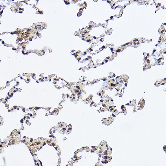
Immunohistochemistry analysis of paraffin-embedded Rat lung using DDX3X Rabbit pAb (A5637) at dilution of 1:100 (40x lens). High pressure antigen retrieval performed with 0.01M Citrate buffer (pH 6.0) prior to IHC staining. |
|
|
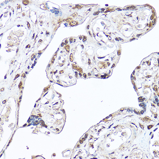
Immunohistochemistry analysis of paraffin-embedded Human placenta using DDX3X Rabbit pAb (A5637) at dilution of 1:100 (40x lens). High pressure antigen retrieval performed with 0.01M Citrate buffer (pH 6.0) prior to IHC staining. |
|
|
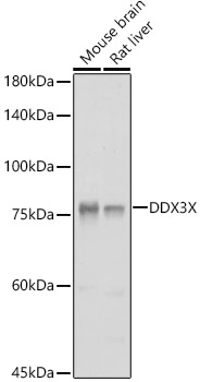
Western blot analysis of various lysates using DDX3X Rabbit pAb (A5637) at 1:1000 dilution. Secondary antibody: HRP-conjugated Goat anti-Rabbit IgG (H+L) (AS014) at 1:10000 dilution. Lysates/proteins: 25µg per lane. Blocking buffer: 3% nonfat dry milk in TBST. Detection: ECL Basic Kit (RM00020). Exposure time: 1s. |
|
|
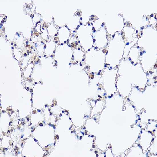
Immunohistochemistry analysis of paraffin-embedded Mouse lung using DDX3X Rabbit pAb (A5637) at dilution of 1:100 (40x lens). High pressure antigen retrieval performed with 0.01M Citrate buffer (pH 6.0) prior to IHC staining. |
|
|
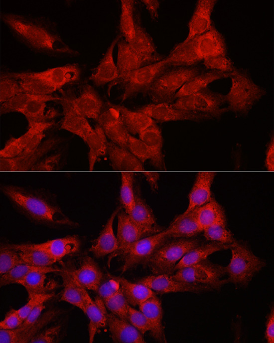
Immunofluorescence analysis of C6 cells using DDX3X Rabbit pAb (A5637) at dilution of 1:100 (40x lens). Secondary antibody: Cy3-conjugated Goat anti-Rabbit IgG (H+L) (AS007) at 1:500 dilution. Blue: DAPI for nuclear staining. |
|
|
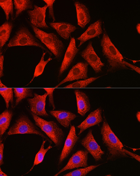
Immunofluorescence analysis of NIH/3T3 cells using DDX3X Rabbit pAb (A5637) at dilution of 1:100 (40x lens). Secondary antibody: Cy3-conjugated Goat anti-Rabbit IgG (H+L) (AS007) at 1:500 dilution. Blue: DAPI for nuclear staining. |
|
|
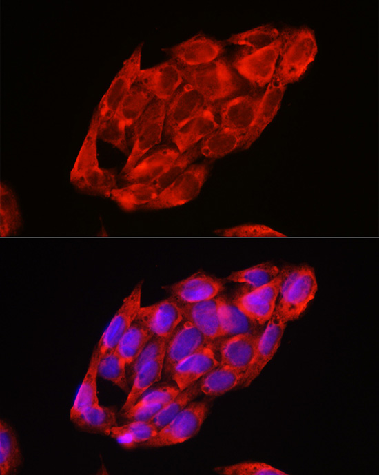
Immunofluorescence analysis of U2OS cells using DDX3X Rabbit pAb (A5637) at dilution of 1:100 (40x lens). Secondary antibody: Cy3-conjugated Goat anti-Rabbit IgG (H+L) (AS007) at 1:500 dilution. Blue: DAPI for nuclear staining. |
|
|
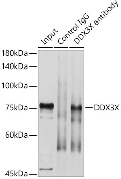
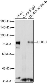
Immunoprecipitation analysis of 600 µg extracts of Mouse brain using 3 µg DDX3X antibody (A5637). Western blot was performed from the immunoprecipitate using DDX3X antibody (A5637) at a dilution of 1:1000. |
|
|
Immunoprecipitation analysis of 300 µg extracts of C6 cells using 3 µg DDX3X antibody (A5637). Western blot was performed from the immunoprecipitate using DDX3X antibody (A5637) at a dilution of 1:3000. |
Product Guarantee and Expert Support
