
UBE2C Rabbit pAb, Unconjugated, Polyclonal
Catalog Number:
ABB-A5499
- Images (8)
| Article Name: | UBE2C Rabbit pAb, Unconjugated, Polyclonal |
| Biozol Catalog Number: | ABB-A5499 |
| Supplier Catalog Number: | A5499 |
| Alternative Catalog Number: | ABB-A5499-100UL,ABB-A5499-20UL,ABB-A5499-1000UL,ABB-A5499-500UL |
| Manufacturer: | ABclonal |
| Host: | Rabbit |
| Category: | Antikörper |
| Application: | ELISA, IF, IHC-P, IP, WB |
| Species Reactivity: | Human |
| Immunogen: | Recombinant protein (or fragment).This information is considered to be commercially sensitive. |
| Conjugation: | Unconjugated |
| Alternative Names: | UBCH10, dJ447F3.2, UBE2C |
| The modification of proteins with ubiquitin is an important cellular mechanism for targeting abnormal or short-lived proteins for degradation. Ubiquitination involves at least three classes of enzymes: ubiquitin-activating enzymes, ubiquitin-conjugating enzymes, and ubiquitin-protein ligases. This gene encodes a member of the E2 ubiquitin-conjugating enzyme family. The encoded protein is required for the destruction of mitotic cyclins and for cell cycle progression, and may be involved in cancer progression. Multiple transcript variants encoding different isoforms have been found for this gene. Pseudogenes of this gene have been defined on chromosomes 4, 14, 15, 18, and 19. |
| Clonality: | Polyclonal |
| Molecular Weight: | 20kDa |
| NCBI: | 11065 |
| UniProt: | O00762 |
| Purity: | Affinity purification |
| Sequence: | MASQNRDPAATSVAAARKGAEPSGGAARGPVGKRLQQELMTLMMSGDKGISAFPESDNLFKWVGTIHGAAGTVYEDLRYKLSLEFPSGYPYNAPTVKFLTPCYHPNVDTQGNICLDILKEKWSALYDVRTILLSIQSLLGEPNIDSPLNTHAAELWKNPTAFKKYLQETYSKQVTSQEP |
| Target: | UBE2C |
| Antibody Type: | Primary Antibody |
| Application Dilute: | WB,1:100 - 1:500|IHC-P,1:50 - 1:200|IF/ICC,1:50 - 1:200|IP,0.5µg-4µg antibody for 200µg-400µg extracts of whole cells|ELISA,Recommended starting concentration is 1 µg/mL. Please optimize the concentration based on your specific assay requirements. |
| Application Notes: | Cross-Reactivity: Human,Mouse,Rat. ResearchArea: Epigenetics Nuclear Signaling,Cell Biology Developmental Biology,Apoptosis,Ubiquitin,Ubiquitin-Proteasome Signaling Pathway. Shipping: Ice Bag |
|
|
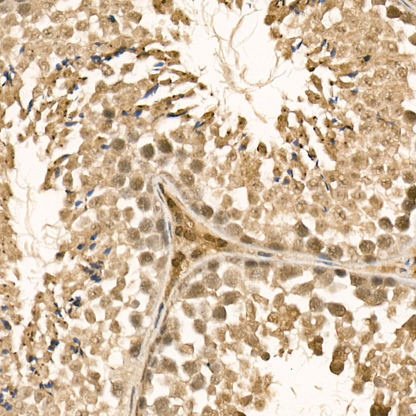
Immunohistochemistry analysis of paraffin-embedded Mouse testis using UBE2C Rabbit pAb (A5499) at dilution of 1:100 (40x lens). High pressure antigen retrieval performed with 0.01M Citrate buffer (pH 6.0) prior to IHC staining. |
|
|
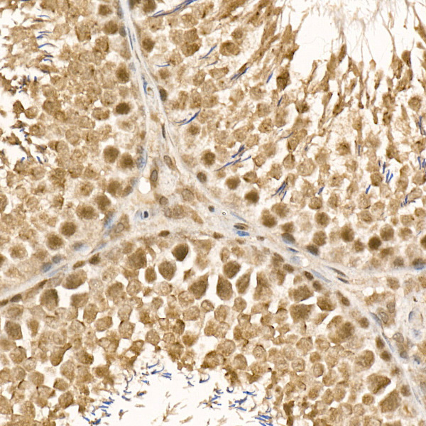
Immunohistochemistry analysis of paraffin-embedded Rat testis using UBE2C Rabbit pAb (A5499) at dilution of 1:100 (40x lens). High pressure antigen retrieval performed with 0.01M Citrate buffer (pH 6.0) prior to IHC staining. |
|
|
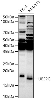
Western blot analysis of various lysates, using UBE2C Rabbit pAb (A5499) at 1:500 dilution. Secondary antibody: HRP-conjugated Goat anti-Rabbit IgG (H+L) (AS014) at 1:10000 dilution. Lysates/proteins: 25µg per lane. Blocking buffer: 3% nonfat dry milk in TBST. Detection: ECL Enhanced Kit (RM00021). Exposure time: 90s. |
|
|
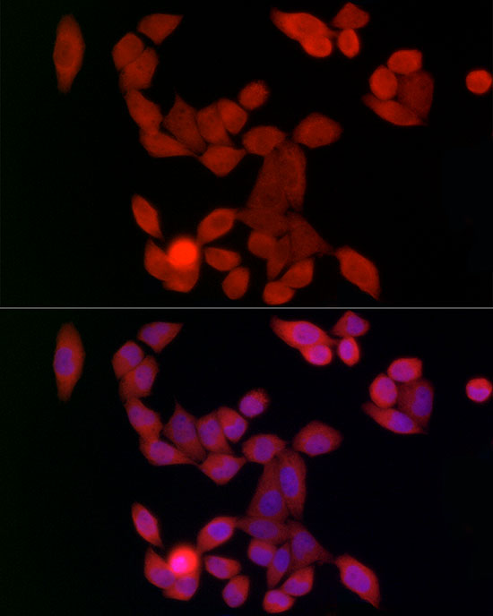
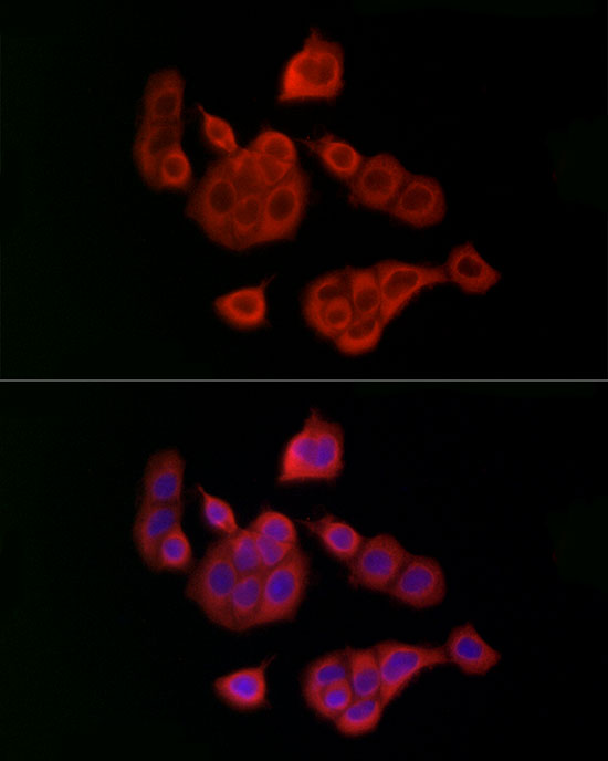
Immunofluorescence analysis of HeLa cells using UBE2C Rabbit pAb (A5499) at dilution of 1:100 (40x lens). Secondary antibody: Cy3-conjugated Goat anti-Rabbit IgG (H+L) (AS007) at 1:500 dilution. Blue: DAPI for nuclear staining. |
|
|
Immunofluorescence analysis of MCF7 cells using UBE2C Rabbit pAb (A5499) at dilution of 1:100 (40x lens). Secondary antibody: Cy3-conjugated Goat anti-Rabbit IgG (H+L) (AS007) at 1:500 dilution. Blue: DAPI for nuclear staining. |
|
|
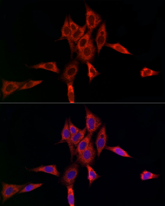
Immunofluorescence analysis of NIH/3T3 cells using UBE2C Rabbit pAb (A5499) at dilution of 1:100 (40x lens). Secondary antibody: Cy3-conjugated Goat anti-Rabbit IgG (H+L) (AS007) at 1:500 dilution. Blue: DAPI for nuclear staining. |
|
|
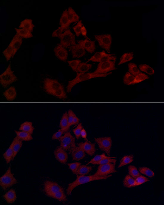
Immunofluorescence analysis of PC-12 cells using UBE2C Rabbit pAb (A5499) at dilution of 1:100 (40x lens). Secondary antibody: Cy3-conjugated Goat anti-Rabbit IgG (H+L) (AS007) at 1:500 dilution. Blue: DAPI for nuclear staining. |
|
|
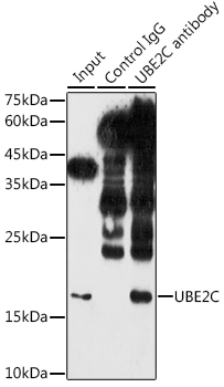
Immunoprecipitation analysis of 300 µg extracts of NIH/3T3 cells using 3 µg UBE2C antibody (A5499). Western blot was performed from the immunoprecipitate using UBE2C antibody (A5499) at a dilution of 1:1000. |
Product Guarantee and Expert Support
