
PAR2 Rabbit mAb, Unconjugated, Monoclonal
Catalog Number:
ABB-A5103
- Images (9)

| Article Name: | PAR2 Rabbit mAb, Unconjugated, Monoclonal |
| Biozol Catalog Number: | ABB-A5103 |
| Supplier Catalog Number: | A5103 |
| Alternative Catalog Number: | ABB-A5103-20UL,ABB-A5103-100UL,ABB-A5103-1000UL,ABB-A5103-500UL |
| Manufacturer: | ABclonal |
| Host: | Rabbit |
| Category: | Antikörper |
| Application: | ELISA, IHC-P, WB |
| Species Reactivity: | Human |
| Immunogen: | Synthetic peptide. This information is considered to be commercially sensitive. |
| Conjugation: | Unconjugated |
| Alternative Names: | PAR2, GPR11 |
| This gene encodes a member of the G-protein coupled receptor 1 family of proteins. The encoded cell surface receptor is activated through proteolytic cleavage of its extracellular amino terminus, resulting in a new amino terminus that acts as a tethered ligand that binds to an extracellular loop domain. Activation of the receptor has been shown to stimulate vascular smooth muscle relaxation, dilate blood vessels, increase blood flow, and lower blood pressure. This protein is also important in the inflammatory response, as well as innate and adaptive immunity. |
| Application Dilute: | WB,1:500 - 1:2000|IHC-P,1:500 - 1:2000|ELISA,Recommended starting concentration is 1 µg/mL. Please optimize the concentration based on your specific assay requirements. |
| Application Notes: | Cross-Reactivity: Human,Mouse,Rat. ResearchArea: Signal Transduction,G protein signaling,G-Protein-Coupled ReceptorsGPCR. Shipping: Ice Bag |
|
|
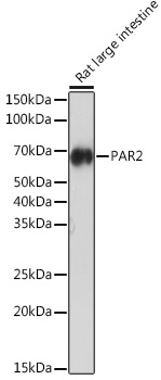
Western blot analysis of lysates from Rat large intestine, using PAR2 Rabbit mAb (A5103) at 1:1000 dilution. Secondary antibody: HRP-conjugated Goat anti-Rabbit IgG (H+L) (AS014) at 1:10000 dilution. Lysates/proteins: 25µg per lane. Blocking buffer: 3% nonfat dry milk in TBST. Detection: ECL Enhanced Kit (RM00021). Exposure time: 3min. |
|
|
Immunohistochemistry analysis of paraffin-embedded Human breast tissue using PAR2 Rabbit mAb (A5103) at a dilution of 1:500 (40x lens). High pressure antigen retrieval performed with 0.01M Tris-EDTA Buffer (pH 9.0) prior to IHC staining. |
|
|
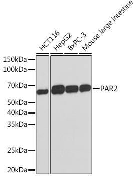
Western blot analysis of various lysates using PAR2 Rabbit mAb (A5103) at 1:1000 dilution. Secondary antibody: HRP-conjugated Goat anti-Rabbit IgG (H+L) (AS014) at 1:10000 dilution. Lysates/proteins: 25µg per lane. Blocking buffer: 3% nonfat dry milk in TBST. Detection: ECL Basic Kit (RM00020). Exposure time: 3min. |
|
|
Immunohistochemistry analysis of paraffin-embedded Human breast cancer tissue using PAR2 Rabbit mAb (A5103) at a dilution of 1:500 (40x lens). High pressure antigen retrieval performed with 0.01M Tris-EDTA Buffer (pH 9.0) prior to IHC staining. |
|
|
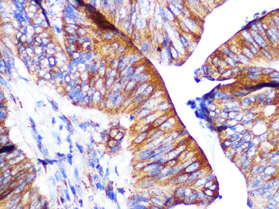
Immunohistochemistry analysis of paraffin-embedded Human colon carcinoma tissue using PAR2 Rabbit mAb (A5103) at a dilution of 1:500 (40x lens). High pressure antigen retrieval performed with 0.01M Tris-EDTA Buffer (pH 9.0) prior to IHC staining. |
|
|
Immunohistochemistry analysis of paraffin-embedded Human colon tissue using PAR2 Rabbit mAb (A5103) at a dilution of 1:500 (40x lens). High pressure antigen retrieval performed with 0.01M Tris-EDTA Buffer (pH 9.0) prior to IHC staining. |
|
|
Immunohistochemistry analysis of paraffin-embedded Human gastric cancer tissue using PAR2 Rabbit mAb (A5103) at a dilution of 1:500 (40x lens). High pressure antigen retrieval performed with 0.01M Tris-EDTA Buffer (pH 9.0) prior to IHC staining. |
|
|
Immunohistochemistry analysis of paraffin-embedded Human pancreatic cancer tissue using PAR2 Rabbit mAb (A5103) at a dilution of 1:500 (40x lens). High pressure antigen retrieval performed with 0.01M Tris-EDTA Buffer (pH 9.0) prior to IHC staining. |
|
|
Immunohistochemistry analysis of paraffin-embedded Human spleen tissue using PAR2 Rabbit mAb (A5103) at a dilution of 1:500 (40x lens). High pressure antigen retrieval performed with 0.01M Tris-EDTA Buffer (pH 9.0) prior to IHC staining. |
Product Guarantee and Expert Support
