
ASH2L Rabbit mAb, Unconjugated, Monoclonal
Catalog Number:
ABB-A4892
- Images (9)

| Article Name: | ASH2L Rabbit mAb, Unconjugated, Monoclonal |
| Biozol Catalog Number: | ABB-A4892 |
| Supplier Catalog Number: | A4892 |
| Alternative Catalog Number: | ABB-A4892-100UL,ABB-A4892-20UL |
| Manufacturer: | ABclonal |
| Host: | Rabbit |
| Category: | Antikörper |
| Application: | ELISA, IF, IHC-P, WB |
| Species Reactivity: | Human |
| Immunogen: | Synthetic peptide. This information is considered to be commercially sensitive. |
| Conjugation: | Unconjugated |
| Alternative Names: | ASH2, Bre2, ASH2L1, ASH2L2, ASH2L |
| Enables beta-catenin binding activity and transcription cis-regulatory region binding activity. Contributes to histone methyltransferase activity (H3-K4 specific). Involved in histone H3-K4 methylation, positive regulation of cell population proliferation, and response to estrogen. Acts upstream of or within cellular response to DNA damage stimulus. Located in nucleus. Part of MLL3/4 complex and Set1C/COMPASS complex. |
| Application Dilute: | WB,1:500 - 1:1000|IF/ICC,1:50 - 1:200|IF-P,1:50 - 1:200|IHC-P,1:50 - 1:200|ELISA,Recommended starting concentration is 1 µg/mL. Please optimize the concentration based on your specific assay requirements. |
| Application Notes: | Cross-Reactivity: Human,Mouse,Rat. ResearchArea: Epigenetics Nuclear Signaling,Chromatin Remodeling,Cancer. Shipping: Ice Bag |
|
|
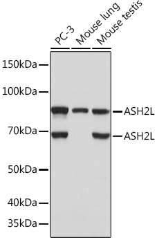
Western blot analysis of various lysates using ASH2L Rabbit mAb (A4892) at 1:1000 dilution. Secondary antibody: HRP-conjugated Goat anti-Rabbit IgG (H+L) (AS014) at 1:10000 dilution. Lysates/proteins: 25µg per lane. Blocking buffer: 3% nonfat dry milk in TBST. Detection: ECL Basic Kit (RM00020). Exposure time: 5s. |
|
|
Immunohistochemistry analysis of paraffin-embeddedMouse lung tissue usingASH2L Rabbit mAb(A4892) at a dilution of 1:200 (40x lens).High pressure antigen retrieval was performed with 0.01 M citrate buffer (pH 6.0) prior to IHC staining. |
|
|
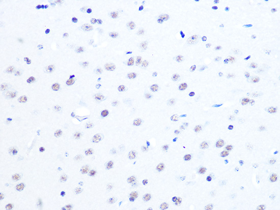
Immunohistochemistry analysis of paraffin-embeddedRat brain tissue usingASH2L Rabbit mAb(A4892) at a dilution of 1:200 (40x lens).High pressure antigen retrieval was performed with 0.01 M citrate buffer (pH 6.0) prior to IHC staining. |
|
|
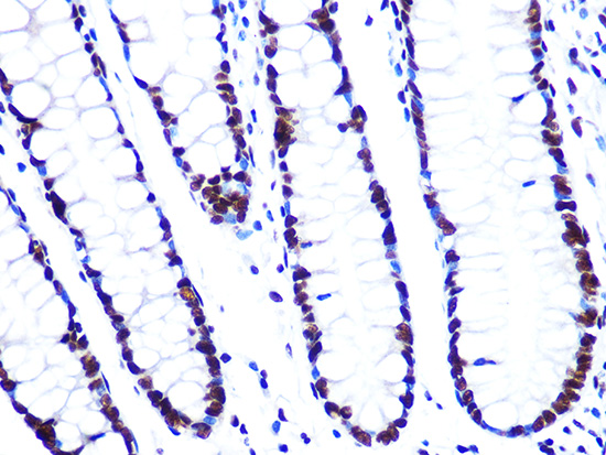
Immunohistochemistry analysis of paraffin-embeddedHuman colon carcinoma tissue usingASH2L Rabbit mAb(A4892) at a dilution of 1:200 (40x lens).High pressure antigen retrieval was performed with 0.01 M citrate buffer (pH 6.0) prior to IHC staining. |
|
|
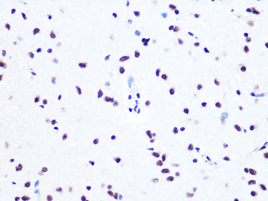
Immunohistochemistry analysis of paraffin-embeddedMouse brain tissue usingASH2L Rabbit mAb(A4892) at a dilution of 1:200 (40x lens).High pressure antigen retrieval was performed with 0.01 M citrate buffer (pH 6.0) prior to IHC staining. |
|
|
Immunohistochemistry analysis of paraffin-embeddedHuman cervix cancer tissue usingASH2L Rabbit mAb(A4892) at a dilution of 1:200 (40x lens).High pressure antigen retrieval was performed with 0.01 M citrate buffer (pH 6.0) prior to IHC staining. |
|
|
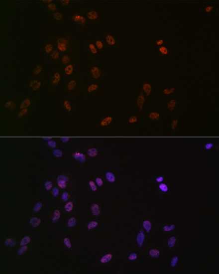
Confocal imaging of C2C12 cells usingASH2L Rabbit mAb (A4892, dilution 1:100) followed by a further incubation with Cy3 Goat Anti-Rabbit IgG (H+L) (AS007, dilution 1:500) (Red). The cells were counterstained with alpha-Tubulin Mouse mAb (AC012, dilution 1:400) followed by incubation with ABflo 488-conjugated Goat Anti-Mouse IgG (H+L) Ab (AS076, dilution 1:500) (Green).DAPI was used for nuclear staining (Blue). Objective: 100x. |
|
|
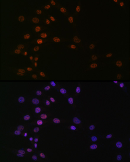
Confocal imaging of NIH/3T3 cells usingASH2L Rabbit mAb (A4892, dilution 1:100) followed by a further incubation with Cy3 Goat Anti-Rabbit IgG (H+L) (AS007, dilution 1:500) (Red). The cells were counterstained with alpha-Tubulin Mouse mAb (AC012, dilution 1:400) followed by incubation with ABflo 488-conjugated Goat Anti-Mouse IgG (H+L) Ab (AS076, dilution 1:500) (Green). DAPI was used for nuclear staining (Blue). Objective: 100x. |
|
|
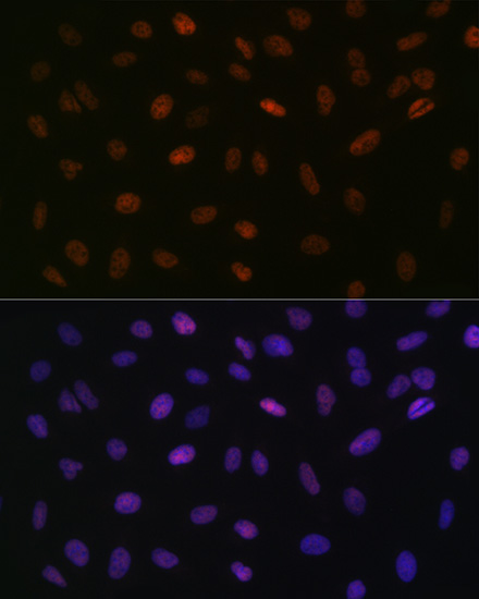
Confocal imaging ofparaffin-embedded Mouse colon tissue usingASH2L Rabbit mAb (A4892, dilution 1:100) followed by a further incubation with Cy3 Goat Anti-Rabbit IgG (H+L) (AS007, dilution 1:500) (Red). DAPI was used for nuclear staining (Blue). Objective: 40x. Perform high pressure antigen retrieval with 0.01 M citrate buffer (pH 6.0) prior to IF staining. |
Product Guarantee and Expert Support
