
Cytokeratin 7 (KRT7) Rabbit mAb, Unconjugated, Monoclonal
Catalog Number:
ABB-A4765
- Images (9)

| Article Name: | Cytokeratin 7 (KRT7) Rabbit mAb, Unconjugated, Monoclonal |
| Biozol Catalog Number: | ABB-A4765 |
| Supplier Catalog Number: | A4765 |
| Alternative Catalog Number: | ABB-A4765-100UL,ABB-A4765-20UL |
| Manufacturer: | ABclonal |
| Host: | Rabbit |
| Category: | Antikörper |
| Application: | ELISA, IF, IHC-P, WB |
| Species Reactivity: | Human |
| Immunogen: | Synthetic peptide. This information is considered to be commercially sensitive. |
| Conjugation: | Unconjugated |
| Alternative Names: | K7, CK7, SCL, K2C7, Cytokeratin 7 (KRT7) |
| The protein encoded by this gene is a member of the keratin gene family. The type II cytokeratins consist of basic or neutral proteins which are arranged in pairs of heterotypic keratin chains coexpressed during differentiation of simple and stratified epithelial tissues. This type II cytokeratin is specifically expressed in the simple epithelia lining the cavities of the internal organs and in the gland ducts and blood vessels. The genes encoding the type II cytokeratins are clustered in a region of chromosome 12q12-q13. Alternative splicing may result in several transcript variants, however, not all variants have been fully described. |
| Application Dilute: | WB,1:1000 - 1:4000|IF-P,1:50 - 1:200|IHC-P,1:500 - 1:2000|ELISA,Recommended starting concentration is 1 µg/mL. Please optimize the concentration based on your specific assay requirements. |
| Application Notes: | Cross-Reactivity: Human,Mouse,Rat. ResearchArea: Signal Transduction,Cell Biology Developmental Biology,Cytoskeleton,Intermediate Filaments,Extracellular Matrix,Keratin. Shipping: Ice Bag |
|
|
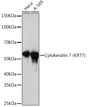
Western blot analysis of various lysates using Cytokeratin 7 (KRT7) (KRT7) Rabbit mAb (A4765) at 1:1000 dilution. Secondary antibody: HRP-conjugated Goat anti-Rabbit IgG (H+L) (AS014) at 1:10000 dilution. Lysates/proteins: 25µg per lane. Blocking buffer: 3% nonfat dry milk in TBST. Detection: ECL Basic Kit (RM00020). Exposure time: 10s. |
|
|
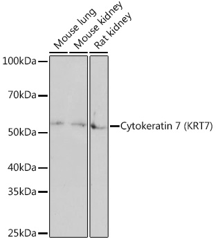
Western blot analysis of various lysates using Cytokeratin 7 (KRT7) (KRT7) Rabbit mAb (A4765) at 1:1000 dilution. Secondary antibody: HRP-conjugated Goat anti-Rabbit IgG (H+L) (AS014) at 1:10000 dilution. Lysates/proteins: 25µg per lane. Blocking buffer: 3% nonfat dry milk in TBST. Detection: ECL Basic Kit (RM00020). Exposure time: 3min. |
|
|
Immunohistochemistry analysis of paraffin-embedded Human liver tissue using Cytokeratin 7 (KRT7) Rabbit mAb (A4765) at a dilution of 1:1000 (40x lens). High pressure antigen retrieval performed with 0.01M Tris-EDTA Buffer (pH 9.0) prior to IHC staining. |
|
|
Immunohistochemistry analysis of paraffin-embedded Human kidney tissue using Cytokeratin 7 (KRT7) Rabbit mAb (A4765) at a dilution of 1:1000 (40x lens). High pressure antigen retrieval performed with 0.01M Tris-EDTA Buffer (pH 9.0) prior to IHC staining. |
|
|
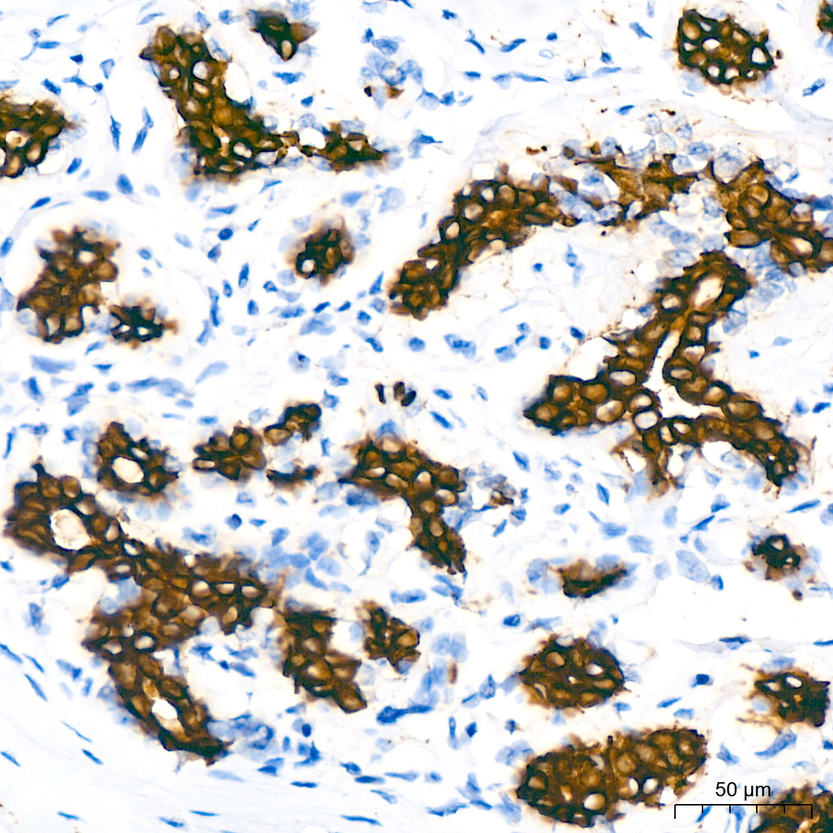
Immunohistochemistry analysis of paraffin-embedded Human lung cancer tissue using Cytokeratin 7 (KRT7) Rabbit mAb (A4765) at a dilution of 1:1000 (40x lens). High pressure antigen retrieval performed with 0.01M Tris-EDTA Buffer (pH 9.0) prior to IHC staining. |
|
|
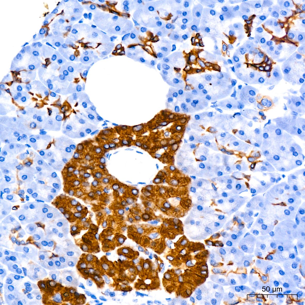
Immunohistochemistry analysis of paraffin-embedded Human pancreas tissue using Cytokeratin 7 (KRT7) Rabbit mAb (A4765) at a dilution of 1:1000 (40x lens). High pressure antigen retrieval performed with 0.01M Tris-EDTA Buffer (pH 9.0) prior to IHC staining. |
|
|
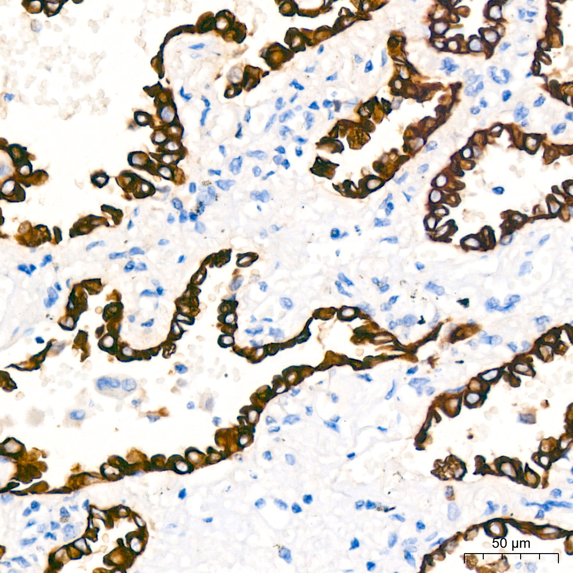
Immunohistochemistry analysis of paraffin-embedded Human placenta tissue using Cytokeratin 7 (KRT7) Rabbit mAb (A4765) at a dilution of 1:1000 (40x lens). High pressure antigen retrieval performed with 0.01M Tris-EDTA Buffer (pH 9.0) prior to IHC staining. |
|
|
Immunohistochemistry analysis of paraffin-embedded Human tonsil tissue using Cytokeratin 7 (KRT7) Rabbit mAb (A4765) at a dilution of 1:1000 (40x lens). High pressure antigen retrieval performed with 0.01M Tris-EDTA Buffer (pH 9.0) prior to IHC staining. |
|
|
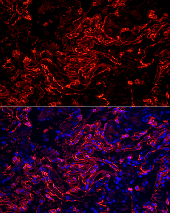
Immunofluorescence analysis of paraffin-embedded human lung using Cytokeratin 7 (KRT7) Rabbit mAb (A4765) at dilution of 1:50 (40x lens). Secondary antibody: Cy3-conjugated Goat anti-Rabbit IgG (H+L) (AS007) at 1:500 dilution. Blue: DAPI for nuclear staining. |
Product Guarantee and Expert Support
