
PP1 beta Rabbit mAb, Unconjugated, Monoclonal
Catalog Number:
ABB-A4364
- Images (8)

| Article Name: | PP1 beta Rabbit mAb, Unconjugated, Monoclonal |
| Biozol Catalog Number: | ABB-A4364 |
| Supplier Catalog Number: | A4364 |
| Alternative Catalog Number: | ABB-A4364-100UL,ABB-A4364-20UL |
| Manufacturer: | ABclonal |
| Host: | Rabbit |
| Category: | Antikörper |
| Application: | ELISA, IF, IHC-P, WB |
| Species Reactivity: | Human |
| Immunogen: | Synthetic peptide. This information is considered to be commercially sensitive. |
| Conjugation: | Unconjugated |
| Alternative Names: | MP, PP1B, PP1c, NSLH2, PP-1B, PPP1CD, PP1beta, PPP1beta, HEL-S-80p, PP1 beta |
| The protein encoded by this gene is one of the three catalytic subunits of protein phosphatase 1 (PP1). PP1 is a serine/threonine specific protein phosphatase known to be involved in the regulation of a variety of cellular processes, such as cell division, glycogen metabolism, muscle contractility, protein synthesis, and HIV-1 viral transcription. Mouse studies suggest that PP1 functions as a suppressor of learning and memory. Two alternatively spliced transcript variants encoding distinct isoforms have been observed. |
| Application Dilute: | WB,1:1000 - 1:6000|IHC-P,1:500 - 1:1000|IF/ICC,1:50 - 1:200|ELISA,Recommended starting concentration is 1 µg/mL. Please optimize the concentration based on your specific assay requirements. |
| Application Notes: | Cross-Reactivity: Human,Mouse,Rat. ResearchArea: Epigenetics Nuclear Signaling,Translation Control,Cancer,Signal Transduction,MAPK-Erk Signaling Pathway,Endocrine Metabolism,Lipid Metabolism,Insulin Receptor Signaling Pathway,Neuroscience,Neurodegenerative Diseases,Dopamine Signaling in Parkinsons Disease. Shipping: Ice Bag |
|
|
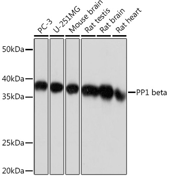
Western blot analysis of various lysates using PP1 beta Rabbit mAb (A4364) at 1:1000 dilution. Secondary antibody: HRP-conjugated Goat anti-Rabbit IgG (H+L) (AS014) at 1:10000 dilution. Lysates/proteins: 25µg per lane. Blocking buffer: 3% nonfat dry milk in TBST. Detection: ECL Basic Kit (RM00020). Exposure time: 1s. |
|
|
Immunohistochemistry analysis of paraffin-embeddedRat colon tissue usingPP1 beta Rabbit mAb(A4364) at a dilution of 1:1000 (40x lens).High pressure antigen retrieval was performed with 0.01 M citrate buffer (pH 6.0) prior to IHC staining. |
|
|
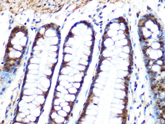
Immunohistochemistry analysis of paraffin-embeddedMouse colon tissue usingPP1 beta Rabbit mAb(A4364) at a dilution of 1:1000 (40x lens).High pressure antigen retrieval was performed with 0.01 M citrate buffer (pH 6.0) prior to IHC staining. |
|
|
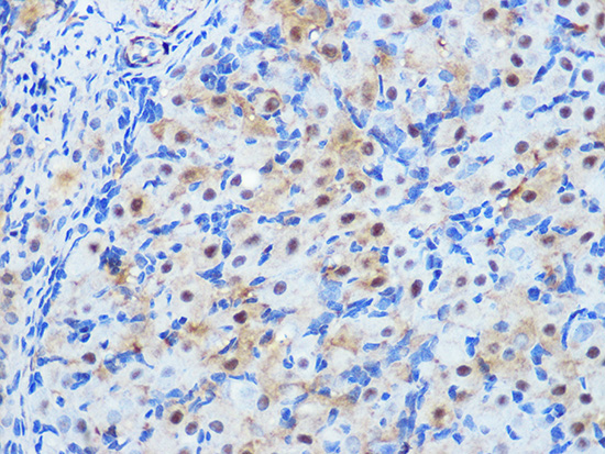
Immunohistochemistry analysis of paraffin-embeddedHuman breast cancer tissue usingPP1 beta Rabbit mAb(A4364) at a dilution of 1:1000 (40x lens).High pressure antigen retrieval was performed with 0.01 M citrate buffer (pH 6.0) prior to IHC staining. |
|
|
Immunohistochemistry analysis of paraffin-embeddedHuman esophagus tissue usingPP1 beta Rabbit mAb(A4364) at a dilution of 1:1000 (40x lens).High pressure antigen retrieval was performed with 0.01 M citrate buffer (pH 6.0) prior to IHC staining. |
|
|
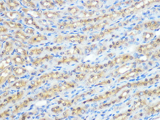
Immunohistochemistry analysis of paraffin-embeddedHuman pancreas tissue usingPP1 beta Rabbit mAb(A4364) at a dilution of 1:1000 (40x lens).High pressure antigen retrieval was performed with 0.01 M citrate buffer (pH 6.0) prior to IHC staining. |
|
|
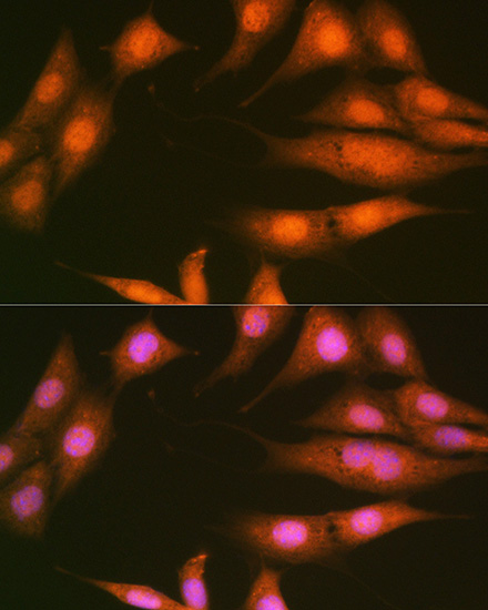
Immunofluorescence analysis of NIH-3T3 cells using PP1 beta Rabbit mAb (A4364) at dilution of 1:100 (40x lens). Secondary antibody: Cy3-conjugated Goat anti-Rabbit IgG (H+L) (AS007) at 1:500 dilution. Blue: DAPI for nuclear staining. |
|
|
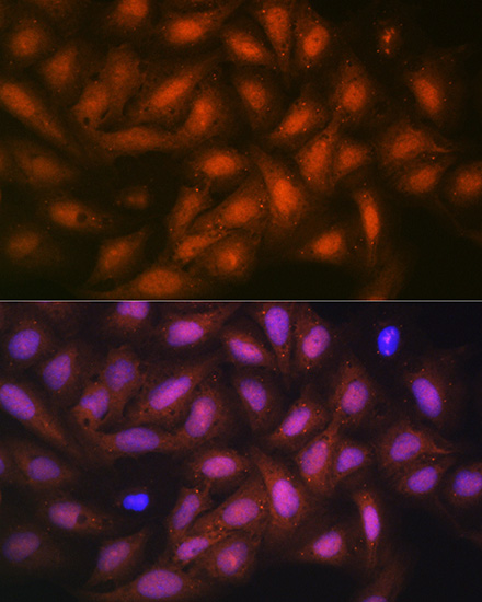
Immunofluorescence analysis of U-2 OS cells using PP1 beta Rabbit mAb (A4364) at dilution of 1:100 (40x lens). Secondary antibody: Cy3-conjugated Goat anti-Rabbit IgG (H+L) (AS007) at 1:500 dilution. Blue: DAPI for nuclear staining. |
Product Guarantee and Expert Support
