
[KO Validated] HMGB1 Rabbit pAb, Unconjugated, Polyclonal
Catalog Number:
ABB-A2553
- Images (8)


| Article Name: | [KO Validated] HMGB1 Rabbit pAb, Unconjugated, Polyclonal |
| Biozol Catalog Number: | ABB-A2553 |
| Supplier Catalog Number: | A2553 |
| Alternative Catalog Number: | ABB-A2553-500UL,ABB-A2553-1000UL,ABB-A2553-20UL,ABB-A2553-100UL |
| Manufacturer: | ABclonal |
| Host: | Rabbit |
| Category: | Antikörper |
| Application: | ELISA, IHC-P, WB |
| Species Reactivity: | Human |
| Immunogen: | Recombinant protein (or fragment).This information is considered to be commercially sensitive. |
| Conjugation: | Unconjugated |
| Alternative Names: | HMG1, HMG3, HMG-1, SBP-1, HMGB1 |
| This gene encodes a protein that belongs to the High Mobility Group-box superfamily. The encoded non-histone, nuclear DNA-binding protein regulates transcription, and is involved in organization of DNA. This protein plays a role in several cellular processes, including inflammation, cell differentiation and tumor cell migration. Multiple pseudogenes of this gene have been identified. Alternative splicing results in multiple transcript variants that encode the same protein. |
| Application Dilute: | WB,1:2000 - 1:6000|IHC-P,1:100 - 1:400|ELISA,Recommended starting concentration is 1 µg/mL. Please optimize the concentration based on your specific assay requirements. |
| Application Notes: | Cross-Reactivity: Human,Mouse,Rat. ResearchArea: Epigenetics Nuclear Signaling,RNA Binding,Cell Biology Developmental Biology,Apoptosis. Shipping: Ice Bag |
|
|
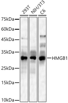
Western blot analysis of lysates from Mouse brain using [KO Validated] HMGB1 Rabbit pAb (A2553) at 1:6000 dilution incubated overnight at 4°C. Secondary antibody: HRP-conjugated Goat anti-Rabbit IgG (H+L) (AS014) at 1:10000 dilution. Lysates/proteins: 25 µg per lane. Blocking buffer: 3% nonfat dry milk in TBST. Detection: ECL Basic Kit (RM00020). Exposure time: 60 s. |
|
|
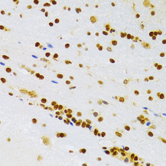
Immunohistochemistry analysis of paraffin-embedded Rat brain using [KO Validated] HMGB1 Rabbit pAb (A2553) at dilution of 1:100 (40x lens). Microwave antigen retrieval performed with 0.01M PBS Buffer (pH 7.2) prior to IHC staining. |
|
|
Western blot analysis of lysates from wild type (WT) and HMGB1 knockout (KO) HeLa cells using [KO Validated] HMGB1 Rabbit pAb (A2553) at 1:6000 dilution incubated overnight at 4°C. Secondary antibody: HRP-conjugated Goat anti-Rabbit IgG (H+L) (AS014) at 1:10000 dilution. Lysates/proteins: 25 µg per lane. Blocking buffer: 3% nonfat dry milk in TBST. Detection: ECL Basic Kit (RM00020). Exposure time: 60 s. |
|
|
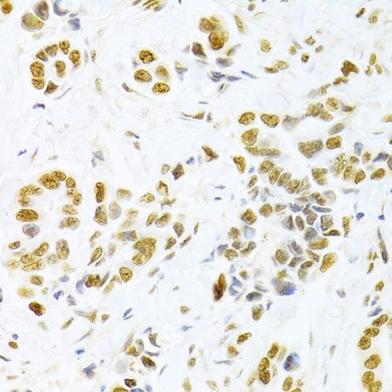
Immunohistochemistry analysis of paraffin-embedded Human breast cancer using [KO Validated] HMGB1 Rabbit pAb (A2553) at dilution of 1:100 (40x lens). Microwave antigen retrieval performed with 0.01M PBS Buffer (pH 7.2) prior to IHC staining. |
|
|
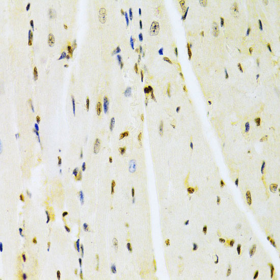
Immunohistochemistry analysis of paraffin-embedded Mouse heart using [KO Validated] HMGB1 Rabbit pAb (A2553) at dilution of 1:100 (40x lens). Microwave antigen retrieval performed with 0.01M PBS Buffer (pH 7.2) prior to IHC staining. |
|
|
Immunohistochemistry analysis of paraffin-embeddedRat liver tissue using[KO Validated] HMGB1 Rabbit pAb(A2553) at a dilution of 1:400 (40x lens).High pressure antigen retrieval was performed with 0.01 M citrate buffer (pH 6.0) prior to IHC staining. |
|
|
Immunohistochemistry analysis of paraffin-embeddedMouse liver tissue using[KO Validated] HMGB1 Rabbit pAb(A2553) at a dilution of 1:400 (40x lens).High pressure antigen retrieval was performed with 0.01 M citrate buffer (pH 6.0) prior to IHC staining. |
|
|
Immunohistochemistry analysis of paraffin-embeddedMouse liver tissue using[KO Validated] HMGB1 Rabbit pAb(A2553) at a dilution of 1:400 (40x lens).High pressure antigen retrieval was performed with 0.01 M citrate buffer (pH 6.0) prior to IHC staining. |
Product Guarantee and Expert Support
