
ABI3 Rabbit mAb, Unconjugated, Monoclonal
Catalog Number:
ABB-A23283
- Images (8)

| Article Name: | ABI3 Rabbit mAb, Unconjugated, Monoclonal |
| Biozol Catalog Number: | ABB-A23283 |
| Supplier Catalog Number: | A23283 |
| Alternative Catalog Number: | ABB-A23283-100UL,ABB-A23283-20UL |
| Manufacturer: | ABclonal |
| Host: | Rabbit |
| Category: | Antikörper |
| Application: | ELISA, IHC-P, WB |
| Species Reactivity: | Human |
| Immunogen: | Recombinant protein (or fragment).This information is considered to be commercially sensitive. |
| Conjugation: | Unconjugated |
| Alternative Names: | NESH, SSH3BP3, ABI3 |
| This gene encodes a member of an adaptor protein family. Members of this family encode proteins containing a homeobox homology domain, proline rich region and Src-homology 3 (SH3) domain, and are components of the Abi/WAVE complex which regulates actin polymerization. The encoded protein inhibits ectopic metastasis of tumor cells as well as cell migration. This may be accomplished through interaction with p21-activated kinase. Alternative splicing results in multiple transcript variants. |
| Application Dilute: | WB,1:500 - 1:1000|IHC-P,1:200 - 1:800|ELISA,Recommended starting concentration is 1 µg/mL. Please optimize the concentration based on your specific assay requirements. |
| Application Notes: | Cross-Reactivity: Human,Mouse,Rat. ResearchArea: Epigenetics Nuclear Signaling,Cancer,Tumor suppressors. Shipping: Ice Bag |
|
|
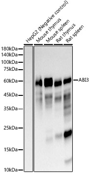
Western blot analysis of various lysates using ABI3 Rabbit mAb (A23283) at 1:1000 dilutionincubated overnight at 4°C. Secondary antibody: HRP-conjugated Goat anti-Rabbit IgG (H+L) (AS014) at 1:10000 dilution. Lysates/proteins: 25 µg per lane. Blocking buffer: 3% nonfat dry milk in TBST. Detection: ECL Basic Kit (RM00020). Negative control (NC): Hep G2 Exposure time: 3s. |
|
|
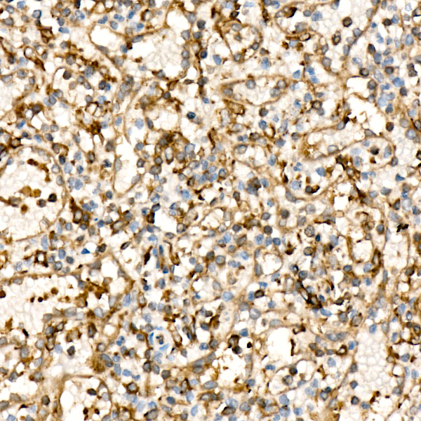
Immunohistochemistry analysis of paraffin-embeddedHuman spleen tissue usingABI3 Rabbit mAb(A23283) at a dilution of 1:200 (40x lens).High pressure antigen retrieval was performed with 0.01 M citrate buffer (pH 6.0) prior to IHC staining. |
|
|
Immunohistochemistry analysis of paraffin-embeddedMouse spleen tissue usingABI3 Rabbit mAb(A23283) at a dilution of 1:200 (40x lens).High pressure antigen retrieval was performed with 0.01 M citrate buffer (pH 6.0) prior to IHC staining. |
|
|
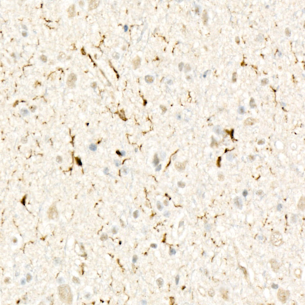
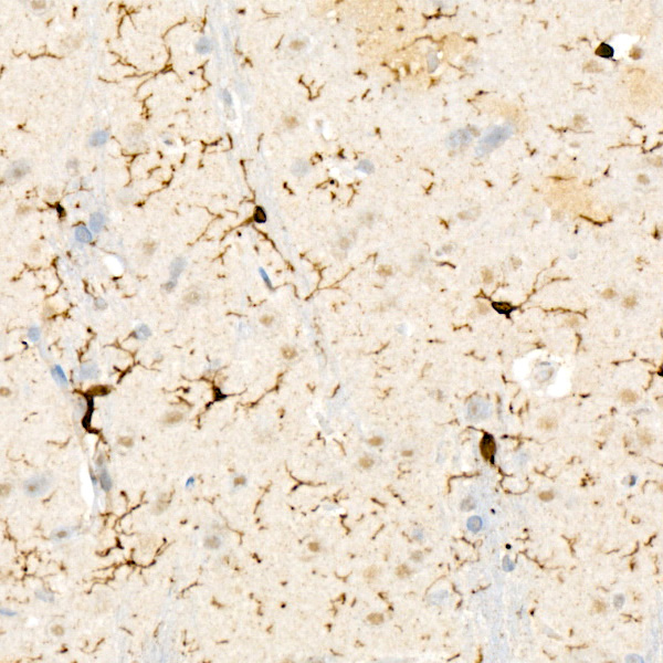
Immunohistochemistry analysis of paraffin-embeddedRat brain tissue usingABI3 Rabbit mAb(A23283) at a dilution of 1:200 (40x lens).High pressure antigen retrieval was performed with 0.01 M citrate buffer (pH 6.0) prior to IHC staining. |
|
|
Immunohistochemistry analysis of paraffin-embeddedMouse colon tissue usingABI3 Rabbit mAb(A23283) at a dilution of 1:200 (40x lens).High pressure antigen retrieval was performed with 0.01 M citrate buffer (pH 6.0) prior to IHC staining. |
|
|
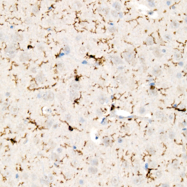
Immunohistochemistry analysis of paraffin-embeddedMouse brain tissue usingABI3 Rabbit mAb(A23283) at a dilution of 1:200 (40x lens).High pressure antigen retrieval was performed with 0.01 M citrate buffer (pH 6.0) prior to IHC staining. |
|
|
Immunohistochemistry analysis of paraffin-embeddedRat spleen tissue usingABI3 Rabbit mAb(A23283) at a dilution of 1:200 (40x lens).High pressure antigen retrieval was performed with 0.01 M citrate buffer (pH 6.0) prior to IHC staining. |
|
|
Immunohistochemistry analysis of paraffin-embeddedHuman small intestine tissue usingABI3 Rabbit mAb(A23283) at a dilution of 1:200 (40x lens).High pressure antigen retrieval was performed with 0.01 M citrate buffer (pH 6.0) prior to IHC staining. |
Product Guarantee and Expert Support
