
USO1 Rabbit mAb, Unconjugated, Monoclonal
Catalog Number:
ABB-A20950
- Images (8)

| Article Name: | USO1 Rabbit mAb, Unconjugated, Monoclonal |
| Biozol Catalog Number: | ABB-A20950 |
| Supplier Catalog Number: | A20950 |
| Alternative Catalog Number: | ABB-A20950-100UL,ABB-A20950-20UL |
| Manufacturer: | ABclonal |
| Host: | Rabbit |
| Category: | Antikörper |
| Application: | ELISA, IF, IHC-P, WB |
| Species Reactivity: | Human |
| Immunogen: | Synthetic peptide. This information is considered to be commercially sensitive. |
| Conjugation: | Unconjugated |
| Alternative Names: | TAP, VDP, P115, USO1 |
| The protein encoded by this gene is a peripheral membrane protein which recycles between the cytosol and the Golgi apparatus during interphase. It is regulated by phosphorylation: dephosphorylated protein associates with the Golgi membrane and dissociates from the membrane upon phosphorylation. Ras-associated protein 1 recruits this protein to coat protein complex II (COPII) vesicles during budding from the endoplasmic reticulum, where it interacts with a set of COPII vesicle-associated SNAREs to form a cis-SNARE complex that promotes targeting to the Golgi apparatus. Alternative splicing results in multiple transcript variants. |
| Application Dilute: | WB,1:500 - 1:1000|IHC-P,1:50 - 1:200|IF/ICC,1:50 - 1:200|ELISA,Recommended starting concentration is 1 µg/mL. Please optimize the concentration based on your specific assay requirements. |
| Application Notes: | Cross-Reactivity: Human,Mouse,Rat. ResearchArea: Epigenetics Nuclear Signaling,RNA Binding,Signal Transduction. Shipping: Ice Bag |
|
|
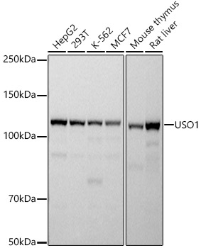
Western blot analysis of various lysates using USO1 Rabbit mAb (A20950) at 1:500 dilution. Secondary antibody: HRP-conjugated Goat anti-Rabbit IgG (H+L) (AS014) at 1:10000 dilution. Lysates/proteins: 25µg per lane. Blocking buffer: 3% nonfat dry milk in TBST. Detection: ECL Basic Kit (RM00020). Exposure time: 1s. |
|
|
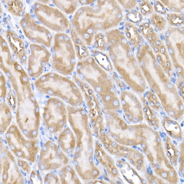
Immunohistochemistry analysis of paraffin-embeddedMouse kidney tissue usingUSO1 Rabbit mAb(A20950) at a dilution of 1:200 (40x lens).High pressure antigen retrieval was performed with 0.01 M citrate buffer (pH 6.0) prior to IHC staining. |
|
|
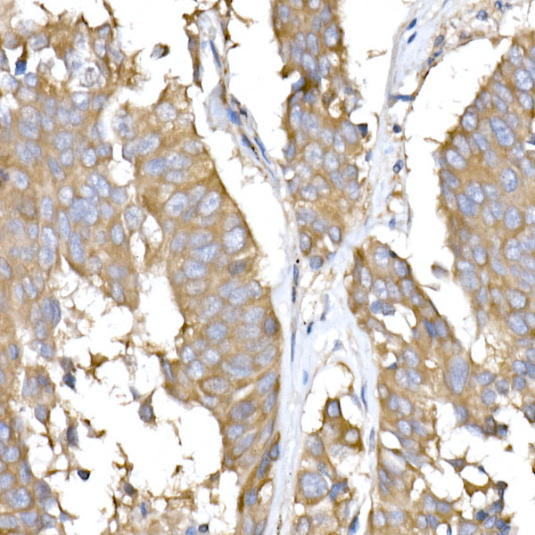
Immunohistochemistry analysis of paraffin-embeddedHuman lung adenocarcinoma tissue usingUSO1 Rabbit mAb(A20950) at a dilution of 1:200 (40x lens).High pressure antigen retrieval was performed with 0.01 M citrate buffer (pH 6.0) prior to IHC staining. |
|
|
Immunohistochemistry analysis of paraffin-embeddedRat kidney tissue usingUSO1 Rabbit mAb(A20950) at a dilution of 1:200 (40x lens).High pressure antigen retrieval was performed with 0.01 M citrate buffer (pH 6.0) prior to IHC staining. |
|
|
Immunohistochemistry analysis of paraffin-embeddedMouse testis tissue usingUSO1 Rabbit mAb(A20950) at a dilution of 1:200 (40x lens).High pressure antigen retrieval was performed with 0.01 M citrate buffer (pH 6.0) prior to IHC staining. |
|
|
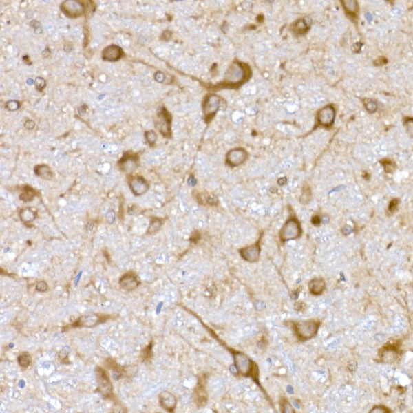
Immunohistochemistry analysis of paraffin-embeddedMouse brain tissue usingUSO1 Rabbit mAb(A20950) at a dilution of 1:200 (40x lens).High pressure antigen retrieval was performed with 0.01 M citrate buffer (pH 6.0) prior to IHC staining. |
|
|
Immunohistochemistry analysis of paraffin-embeddedHuman colon carcinoma tissue usingUSO1 Rabbit mAb(A20950) at a dilution of 1:200 (40x lens).High pressure antigen retrieval was performed with 0.01 M citrate buffer (pH 6.0) prior to IHC staining. |
|
|
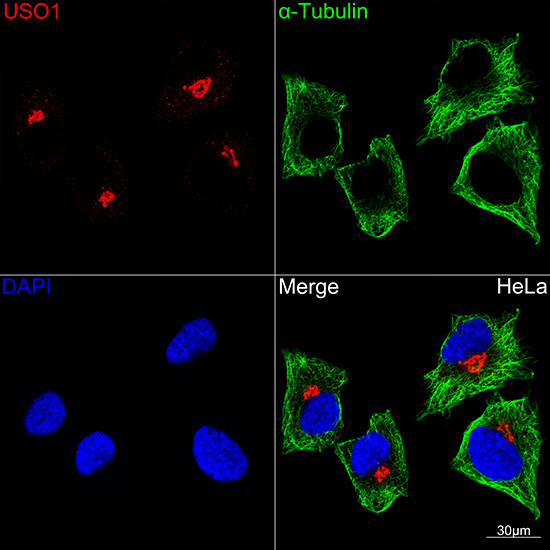
Confocal imaging of HeLa cells using USO1RabbitmAb (A20950, dilution 1:100) (Red). The cells were counterstained with alpha-Tubulin Mouse mAb (AC012, dilution 1:200) (Green). DAPI was used for nuclear staining (blue). Objective: 60x. |
Product Guarantee and Expert Support
