
RBM3 Rabbit mAb, Unconjugated, Monoclonal
Catalog Number:
ABB-A20911
- Images (8)
| Article Name: | RBM3 Rabbit mAb, Unconjugated, Monoclonal |
| Biozol Catalog Number: | ABB-A20911 |
| Supplier Catalog Number: | A20911 |
| Alternative Catalog Number: | ABB-A20911-100UL,ABB-A20911-20UL |
| Manufacturer: | ABclonal |
| Host: | Rabbit |
| Category: | Antikörper |
| Application: | ELISA, IF, IHC-P, WB |
| Species Reactivity: | Human |
| Immunogen: | Synthetic peptide. This information is considered to be commercially sensitive. |
| Conjugation: | Unconjugated |
| Alternative Names: | RNPL, IS1-RNPL, RBM3 |
| This gene is a member of the glycine-rich RNA-binding protein family and encodes a protein with one RNA recognition motif (RRM) domain. Expression of this gene is induced by cold shock and low oxygen tension. A pseudogene exists on chromosome 1. Multiple alternatively spliced transcript variants that are predicted to encode different isoforms have been characterized although some of these variants fit nonsense-mediated decay (NMD) criteria. |
| Application Dilute: | WB,1:500 - 1:1000|IHC-P,1:50 - 1:200|IF/ICC,1:50 - 1:200|ELISA,Recommended starting concentration is 1 µg/mL. Please optimize the concentration based on your specific assay requirements. |
| Application Notes: | Cross-Reactivity: Human,Mouse,Rat. ResearchArea: Epigenetics Nuclear Signaling,RNA Binding,Cancer,Invasion and Metastasis,Endocrine Metabolism. Shipping: Ice Bag |
|
|
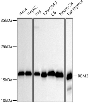
Western blot analysis of various lysates using RBM3 Rabbit mAb (A20911) at 1:500 dilution. Secondary antibody: HRP-conjugated Goat anti-Rabbit IgG (H+L) (AS014) at 1:10000 dilution. Lysates/proteins: 25µg per lane. Blocking buffer: 3% nonfat dry milk in TBST. Detection: ECL Basic Kit (RM00020). Exposure time: 30s. |
|
|
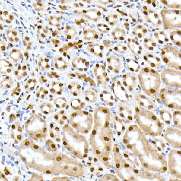
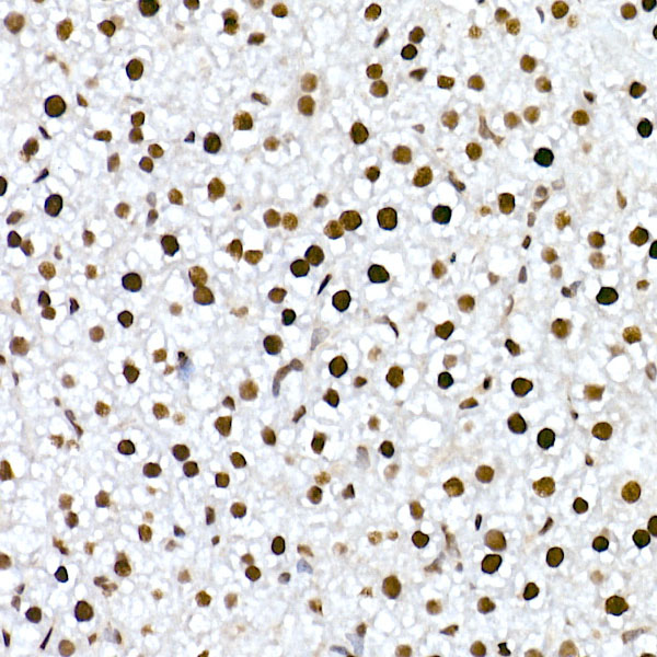
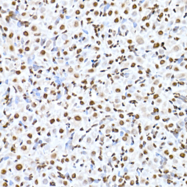
Immunohistochemistry analysis of paraffin-embeddedRat colon tissue usingRBM3 Rabbit mAb(A20911) at a dilution of 1:200 (40x lens).High pressure antigen retrieval was performed with 0.01 M citrate buffer (pH 6.0) prior to IHC staining. |
|
|
Immunohistochemistry analysis of paraffin-embeddedMouse colon tissue usingRBM3 Rabbit mAb(A20911) at a dilution of 1:200 (40x lens).High pressure antigen retrieval was performed with 0.01 M citrate buffer (pH 6.0) prior to IHC staining. |
|
|
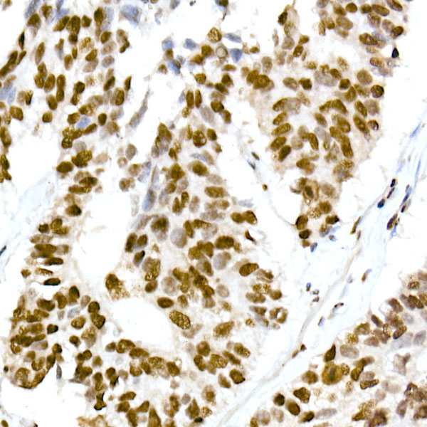
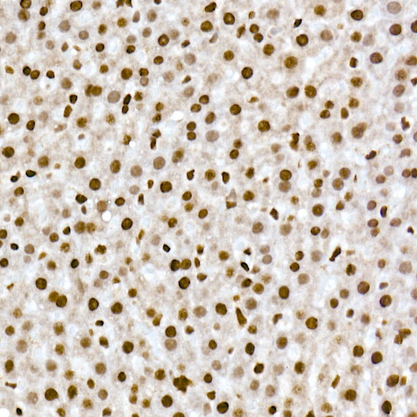
Immunohistochemistry analysis of paraffin-embeddedHuman thyroid tissue usingRBM3 Rabbit mAb(A20911) at a dilution of 1:200 (40x lens).High pressure antigen retrieval was performed with 0.01 M citrate buffer (pH 6.0) prior to IHC staining. |
|
|
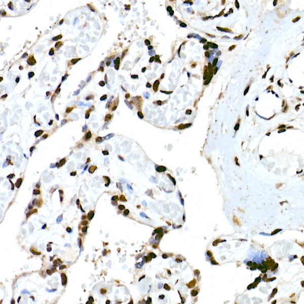
Immunohistochemistry analysis of paraffin-embeddedHuman breast tissue usingRBM3 Rabbit mAb(A20911) at a dilution of 1:200 (40x lens).High pressure antigen retrieval was performed with 0.01 M citrate buffer (pH 6.0) prior to IHC staining. |
|
|
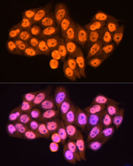
Immunofluorescence analysis of HeLa cells using RBM3 Rabbit mAb (A20911) at dilution of 1:100 (40x lens). Secondary antibody: Cy3-conjugated Goat anti-Rabbit IgG (H+L) (AS007) at 1:500 dilution. Blue: DAPI for nuclear staining. |
|
|
Immunofluorescence analysis of NIH/3T3 cells using RBM3 Rabbit mAb (A20911) at dilution of 1:100 (40x lens). Secondary antibody: Cy3-conjugated Goat anti-Rabbit IgG (H+L) (AS007) at 1:500 dilution. Blue: DAPI for nuclear staining. |
|
|
Immunofluorescence analysis of PC-12 cells using RBM3 Rabbit mAb (A20911) at dilution of 1:100 (40x lens). Secondary antibody: Cy3-conjugated Goat anti-Rabbit IgG (H+L) (AS007) at 1:500 dilution. Blue: DAPI for nuclear staining. |
Product Guarantee and Expert Support
