
LDL Receptor (LDLR) Rabbit mAb, Unconjugated, Monoclonal
Catalog Number:
ABB-A20808
- Images (8)

| Article Name: | LDL Receptor (LDLR) Rabbit mAb, Unconjugated, Monoclonal |
| Biozol Catalog Number: | ABB-A20808 |
| Supplier Catalog Number: | A20808 |
| Alternative Catalog Number: | ABB-A20808-20UL,ABB-A20808-100UL,ABB-A20808-500UL,ABB-A20808-1000UL |
| Manufacturer: | ABclonal |
| Host: | Rabbit |
| Category: | Antikörper |
| Application: | ELISA, IF, IHC-P, WB |
| Species Reactivity: | Human |
| Immunogen: | Synthetic peptide. This information is considered to be commercially sensitive. |
| Conjugation: | Unconjugated |
| Alternative Names: | FH, FHC, FHCL1, LDLCQ2, LDL Receptor (LDLR) |
| The low density lipoprotein receptor (LDLR) gene family consists of cell surface proteins involved in receptor-mediated endocytosis of specific ligands. The encoded protein is normally bound at the cell membrane, where it binds low density lipoprotein/cholesterol and is taken into the cell. Lysosomes release the cholesterol, which is made available for repression of microsomal enzyme 3-hydroxy-3-methylglutaryl coenzyme A (HMG CoA) reductase, the rate-limiting step in cholesterol synthesis. At the same time, a reciprocal stimulation of cholesterol ester synthesis takes place. Mutations in this gene cause the autosomal dominant disorder, familial hypercholesterolemia. Alternate splicing results in multiple transcript variants. |
| Application Dilute: | WB,1:1000 - 1:2000|IHC-P,1:400 - 1:1200|IF/ICC,1:200 - 1:400|ELISA,Recommended starting concentration is 1 µg/mL. Please optimize the concentration based on your specific assay requirements. |
| Application Notes: | Cross-Reactivity: Human,Mouse,Rat. ResearchArea: Cancer,Signal Transduction,Endocrine Metabolism,Lipid Metabolism,Cholesterol Metabolism,Cardiovascular,Heart,Lipids,Cardiovascular diseases,Heart disease. Shipping: Ice Bag |
|
|
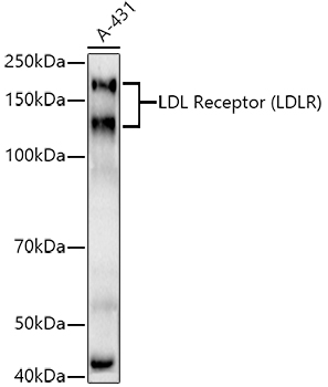
Western blot analysis of lysates from A-431 cells, using LDL Receptor (LDLR) Rabbit mAb (A20808) at1:1000 dilution. Secondary antibody: HRP-conjugated Goat anti-Rabbit IgG (H+L) (AS014) at1:10000 dilution. Lysates/proteins: 25µg per lane. Blocking buffer: 3% nonfat dry milk in TBST. Detection: ECL Basic Kit (RM00020). Exposure time: 180s. |
|
|
Immunohistochemistry analysis of paraffin-embedded Mouse intestin tissue using LDL Receptor (LDLR) Rabbit mAb (A20808) at a dilution of 1:800 (40x lens). High pressure antigen retrieval performed with 0.01M Tris-EDTA Buffer (pH 9.0) prior to IHC staining. |
|
|
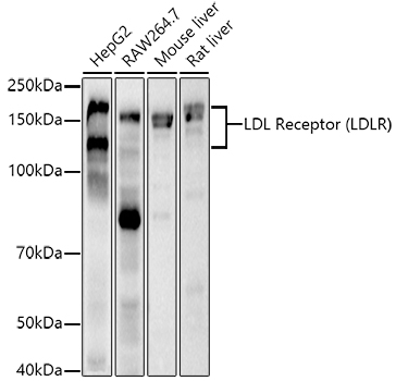
Western blot analysis of various lysates using LDL Receptor (LDLR) Rabbit mAb (A20808) at1:1000 dilution. Secondary antibody: HRP-conjugated Goat anti-Rabbit IgG (H+L) (AS014) at1:10000 dilution. Lysates/proteins: 25µg per lane. Blocking buffer: 3% nonfat dry milk in TBST. Detection: ECL Basic Kit (RM00020). Exposure time: 30s. |
|
|
Immunohistochemistry analysis of paraffin-embedded Mouse liver tissue using LDL Receptor (LDLR) Rabbit mAb (A20808) at a dilution of 1:800 (40x lens). High pressure antigen retrieval performed with 0.01M Tris-EDTA Buffer (pH 9.0) prior to IHC staining. |
|
|
Immunohistochemistry analysis of paraffin-embedded Mouse lung tissue using LDL Receptor (LDLR) Rabbit mAb (A20808) at a dilution of 1:800 (40x lens). High pressure antigen retrieval performed with 0.01M Tris-EDTA Buffer (pH 9.0) prior to IHC staining. |
|
|
Immunohistochemistry analysis of paraffin-embedded Mouse testis tissue using LDL Receptor (LDLR) Rabbit mAb (A20808) at a dilution of 1:800 (40x lens). High pressure antigen retrieval performed with 0.01M Tris-EDTA Buffer (pH 9.0) prior to IHC staining. |
|
|
Immunohistochemistry analysis of paraffin-embedded Rat lung tissue using LDL Receptor (LDLR) Rabbit mAb (A20808) at a dilution of 1:800 (40x lens). High pressure antigen retrieval performed with 0.01M Tris-EDTA Buffer (pH 9.0) prior to IHC staining. |
|
|
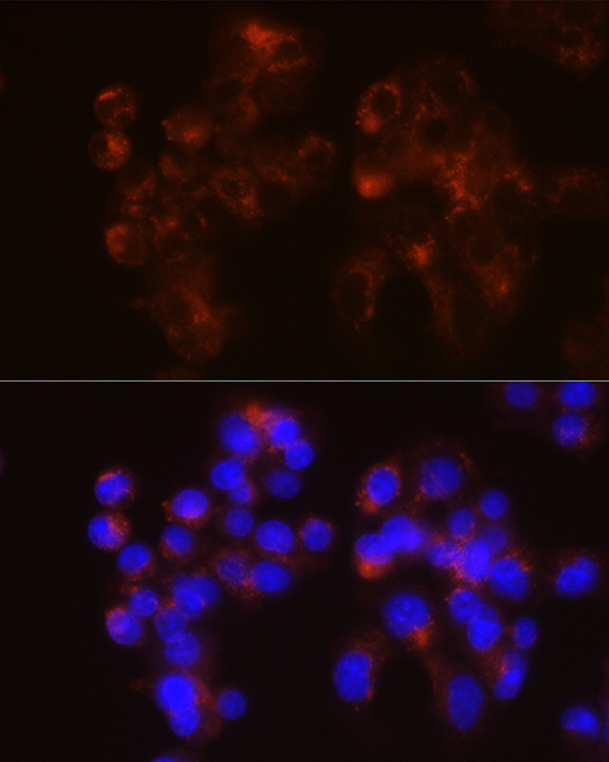
Confocal imaging of Hep G2 cells using LDL Receptor (LDLR) Rabbit mAb (A20808, dilution 1:200) followed by a further incubation with Cy3 Goat Anti-Rabbit IgG (H+L) (AS007, dilution 1:500) (Red). The cells were counterstained with alpha-Tubulin Mouse mAb (AC012, dilution 1:400) followed by incubation with ABflo 488-conjugated Goat Anti-Mouse IgG (H+L) Ab (AS076, dilution 1:500) (Green). DAPI was used for nuclear staining (Blue). Objective: 100x. |
Product Guarantee and Expert Support
