
[KO Validated] SIRT3 Rabbit mAb, Unconjugated, Monoclonal
Catalog Number:
ABB-A20805
- Images (8)

| Article Name: | [KO Validated] SIRT3 Rabbit mAb, Unconjugated, Monoclonal |
| Biozol Catalog Number: | ABB-A20805 |
| Supplier Catalog Number: | A20805 |
| Alternative Catalog Number: | ABB-A20805-100UL,ABB-A20805-20UL,ABB-A20805-1000UL,ABB-A20805-500UL |
| Manufacturer: | ABclonal |
| Host: | Rabbit |
| Category: | Antikörper |
| Application: | ELISA, IHC-P, IP, WB |
| Species Reactivity: | Human |
| Immunogen: | Synthetic peptide. This information is considered to be commercially sensitive. |
| Conjugation: | Unconjugated |
| Alternative Names: | SIR2L3, T3 |
| SIRT3 encodes a member of the sirtuin family of class III histone deacetylases, homologs to the yeast Sir2 protein. The encoded protein is found exclusively in mitochondria, where it can eliminate reactive oxygen species, inhibit apoptosis, and prevent the formation of cancer cells. SIRT3 has far-reaching effects on nuclear gene expression, cancer, cardiovascular disease, neuroprotection, aging, and metabolic control. |
| Application Dilute: | WB,1:1000 - 1:4000|IHC-P,1:500 - 1:2000|IP,0.5µg-4µg antibody for 200µg-400µg extracts of whole cells|ELISA,Recommended starting concentration is 1 µg/mL. Please optimize the concentration based on your specific assay requirements. |
| Application Notes: | Cross-Reactivity: Human,Rat. ResearchArea: Epigenetics Nuclear Signaling. Shipping: Ice Bag |
|
|
Western blot analysis of lysates from wild type (WT) and SIRT3 knockout (KO) 293T cells using [KO Validated] SIRT3 Rabbit mAb (A20805) at 1:1000 dilution incubated overnight at 4°C. Secondary antibody: HRP-conjugated Goat anti-Rabbit IgG (H+L) (AS014) at 1:10000 dilution. Lysates/proteins: 25 µg per lane. Blocking buffer: 3% nonfat dry milk in TBST. Detection: ECL Basic Kit (RM00020). Exposure time: 30s. |
|
|
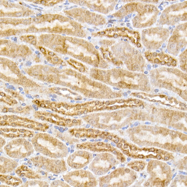
Immunohistochemistry analysis of paraffin-embedded Human kidney tissue using [KO Validated] SIRT3 Rabbit mAb (A20805) at a dilution of 1:2000 (40x lens). High pressure antigen retrieval performed with 0.01M Tris-EDTA Buffer (pH 9.0) prior to IHC staining. |
|
|
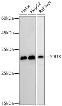
Western blot analysis of various lysates, using [KO Validated] SIRT3 Rabbit mAb (A20805) at 1:1000 dilution. Secondary antibody: HRP-conjugated Goat anti-Rabbit IgG (H+L) (AS014) at 1:10000 dilution. Lysates/proteins: 25µg per lane. Blocking buffer: 3% nonfat dry milk in TBST. Detection: ECL Basic Kit (RM00020). Exposure time: 30s. |
|
|
Immunohistochemistry analysis of paraffin-embedded Human colon carcinoma tissue using [KO Validated] SIRT3 Rabbit mAb (A20805) at a dilution of 1:2000 (40x lens). High pressure antigen retrieval performed with 0.01M Tris-EDTA Buffer (pH 9.0) prior to IHC staining. |
|
|
Immunohistochemistry analysis of paraffin-embedded Human breast cancer tissue using [KO Validated] SIRT3 Rabbit mAb (A20805) at a dilution of 1:2000 (40x lens). High pressure antigen retrieval performed with 0.01M Tris-EDTA Buffer (pH 9.0) prior to IHC staining. |
|
|
Immunohistochemistry analysis of paraffin-embedded Human liver cancer tissue using [KO Validated] SIRT3 Rabbit mAb (A20805) at a dilution of 1:2000 (40x lens). High pressure antigen retrieval performed with 0.01M Tris-EDTA Buffer (pH 9.0) prior to IHC staining. |
|
|
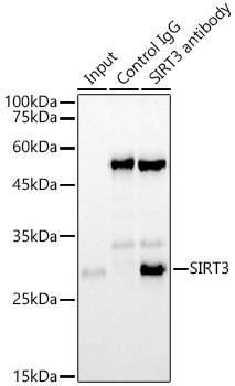
Immunoprecipitation analysis of 300ug extracts of Hela cells using 3ug SIRT3 Rabbit mAb (A20805). Western blot was performed from the immunoprecipitate using SIRT3 Rabbit mAb (A20805) at a dilition of 1:1000. |
|
|
Immunoprecipitation analysis of 300ug extracts of Hela cells using 3ug SIRT3 Rabbit mAb (A20805). Western blot was performed from the immunoprecipitate using SIRT3 Rabbit mAb (A20805) at a dilition of 1:1000. |
Product Guarantee and Expert Support
