
VDAC2 Rabbit pAb, Polyclonal
Catalog Number:
ABB-A18683
- Images (7)
| Article Name: | VDAC2 Rabbit pAb, Polyclonal |
| Biozol Catalog Number: | ABB-A18683 |
| Supplier Catalog Number: | A18683 |
| Alternative Catalog Number: | ABB-A18683-20UL,ABB-A18683-100UL,ABB-A18683-1000UL,ABB-A18683-500UL |
| Manufacturer: | ABclonal |
| Host: | Rabbit |
| Category: | Antikörper |
| Application: | ELISA, IF, IHC-P, WB |
| Species Reactivity: | Human |
| Immunogen: | Synthetic peptide. This information is considered to be commercially sensitive. |
| Alternative Names: | POR, VDAC2 |
| This gene encodes a member of the voltage-dependent anion channel pore-forming family of proteins that are considered the main pathway for metabolite diffusion across the mitochondrial outer membrane. The encoded protein is also thought to be involved in the mitochondrial apoptotic pathway via regulation of BCL2-antagonist/killer 1 protein activity. Pseudogenes have been identified on chromosomes 1, 2, 12 and 21, and alternative splicing results in multiple transcript variants. |
| Application Dilute: | WB,1:500 - 1:1000|IHC-P,1:50 - 1:200|IF/ICC,1:50 - 1:200|ELISA,Recommended starting concentration is 1 µg/mL. Please optimize the concentration based on your specific assay requirements. |
| Application Notes: | Cross-Reactivity: Human,Mouse,Rat. ResearchArea: Cancer,Signal Transduction,Endocrine Metabolism,Mitochondrial metabolism,Mitochondrial markers,Warburg Effect,Neuroscience,Neurodegenerative Diseases. Shipping: Ice Bag |
|
|
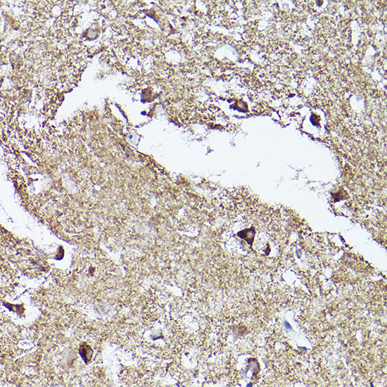
Immunohistochemistry analysis of paraffin-embedded Human brain using VDAC2 Rabbit pAb (A18683) at dilution of 1:100 (40x lens). High pressure antigen retrieval performed with 0.01M Citrate buffer (pH 6.0) prior to IHC staining. |
|
|
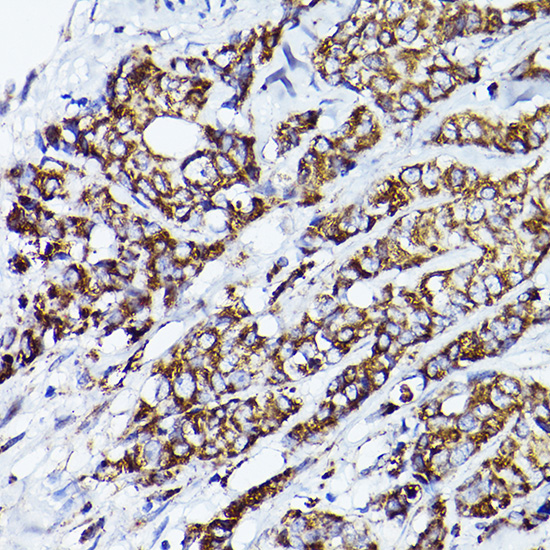
Immunohistochemistry analysis of paraffin-embedded Human breast cancer using VDAC2 Rabbit pAb (A18683) at dilution of 1:100 (40x lens). High pressure antigen retrieval performed with 0.01M Citrate buffer (pH 6.0) prior to IHC staining. |
|
|
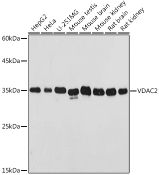
Western blot analysis of various lysates using VDAC2 Rabbit pAb (A18683) at 1:500 dilution. Secondary antibody: HRP-conjugated Goat anti-Rabbit IgG (H+L) (AS014) at 1:10000 dilution. Lysates/proteins: 25µg per lane. Blocking buffer: 3% nonfat dry milk in TBST. Detection: ECL Basic Kit (RM00020). Exposure time: 1s. |
|
|
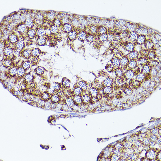
Immunohistochemistry analysis of paraffin-embedded Mouse testis using VDAC2 Rabbit pAb (A18683) at dilution of 1:100 (40x lens). High pressure antigen retrieval performed with 0.01M Citrate buffer (pH 6.0) prior to IHC staining. |
|
|
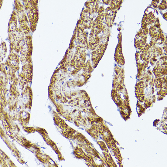
Immunohistochemistry analysis of paraffin-embedded Rat heart using VDAC2 Rabbit pAb (A18683) at dilution of 1:100 (40x lens). High pressure antigen retrieval performed with 0.01M Citrate buffer (pH 6.0) prior to IHC staining. |
|
|
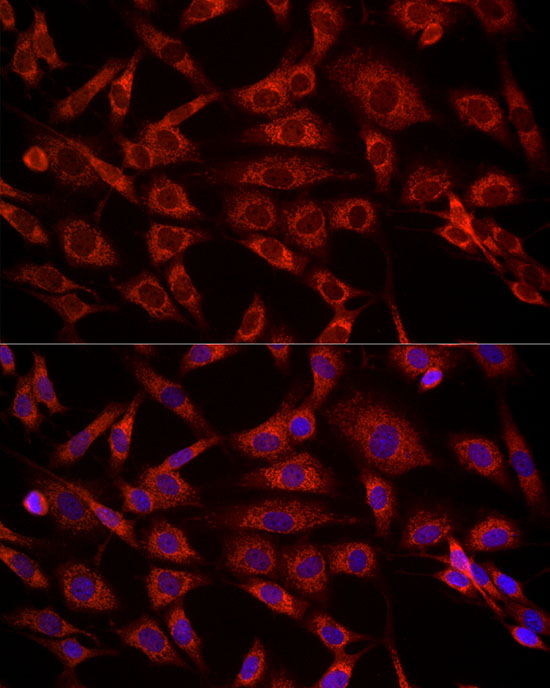
Immunofluorescence analysis of NIH/3T3 cells using VDAC2 Rabbit pAb (A18683) at dilution of 1:100 (40x lens). Secondary antibody: Cy3-conjugated Goat anti-Rabbit IgG (H+L) (AS007) at 1:500 dilution. Blue: DAPI for nuclear staining. |
|
|
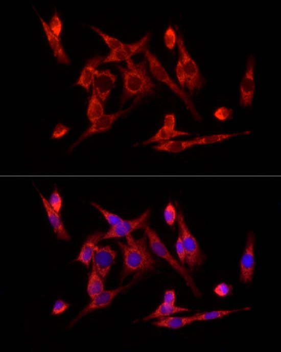
Immunofluorescence analysis of PC-12 cells using VDAC2 Rabbit pAb (A18683) at dilution of 1:100 (40x lens). Secondary antibody: Cy3-conjugated Goat anti-Rabbit IgG (H+L) (AS007) at 1:500 dilution. Blue: DAPI for nuclear staining. |
Product Guarantee and Expert Support
