
IFNAR1 Rabbit pAb, Polyclonal
Catalog Number:
ABB-A18594
- Images (7)

| Article Name: | IFNAR1 Rabbit pAb, Polyclonal |
| Biozol Catalog Number: | ABB-A18594 |
| Supplier Catalog Number: | A18594 |
| Alternative Catalog Number: | ABB-A18594-20UL,ABB-A18594-100UL,ABB-A18594-500UL,ABB-A18594-1000UL |
| Manufacturer: | ABclonal |
| Host: | Rabbit |
| Category: | Antikörper |
| Application: | ELISA, IF, IHC-P, WB |
| Species Reactivity: | Human |
| Immunogen: | Recombinant protein (or fragment).This information is considered to be commercially sensitive. |
| Alternative Names: | AVP, IFRC, IFNAR, IFNBR, IMD106, IFN-alpha-REC, IFNAR1 |
| The protein encoded by this gene is a type I membrane protein that forms one of the two chains of a receptor for interferons alpha and beta. Binding and activation of the receptor stimulates Janus protein kinases, which in turn phosphorylate several proteins, including STAT1 and STAT2. The protein belongs to the type II cytokine receptor family and functions as an antiviral factor. |
| Application Dilute: | WB,1:1000 - 1:5000|IHC-P,1:50 - 1:200|IF/ICC,1:50 - 1:200|ELISA,Recommended starting concentration is 1 µg/mL. Please optimize the concentration based on your specific assay requirements. |
| Application Notes: | Cross-Reactivity: Human,Mouse,Rat. ResearchArea: Epigenetics Nuclear Signaling,Cancer,Tumor immunology,Signal Transduction,Immunology Inflammation,Cytokines,Cell Intrinsic Innate Immunity Signaling Pathway. Shipping: Ice Bag |
|
|
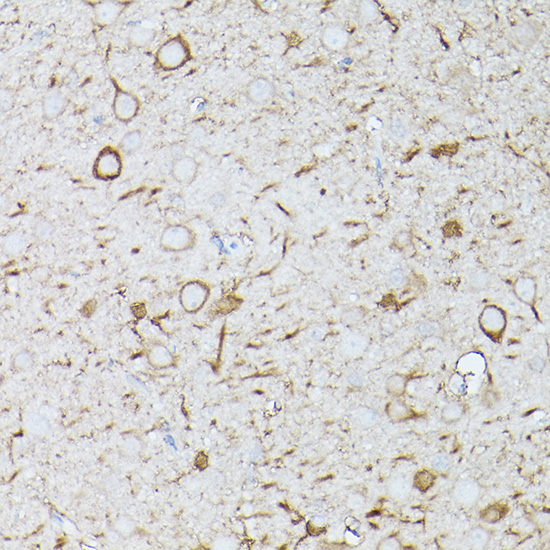
Immunohistochemistry analysis of paraffin-embedded Mouse brain using IFNAR1 Rabbit pAb (A18594) at dilution of 1:100 (40x lens). High pressure antigen retrieval performed with 0.01M Citrate buffer (pH 6.0) prior to IHC staining. |
|
|
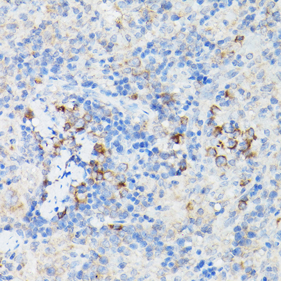
Immunohistochemistry analysis of paraffin-embedded Mouse spleen using IFNAR1 Rabbit pAb (A18594) at dilution of 1:100 (40x lens). High pressure antigen retrieval performed with 0.01M Citrate buffer (pH 6.0) prior to IHC staining. |
|
|
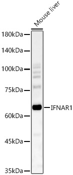
Western blot analysis of various lysates, using IFNAR1 Rabbit pAb (A18594) at 1:2000 dilution. Secondary antibody: HRP-conjugated Goat anti-Rabbit IgG (H+L) (AS014) at 1:10000 dilution. Lysates/proteins: 25µg per lane. Blocking buffer: 3% nonfat dry milk in TBST. Detection: ECL Basic Kit (RM00020). Exposure time: 90s. |
|
|
Immunohistochemistry analysis of paraffin-embedded Human colon tissue using IFNAR1 Rabbit pAb (A18594) at a dilution of 1:200 (40x lens). High pressure antigen retrieval performed with 0.01M Citrate Buffer(pH 6.0) prior to IHC staining. |
|
|
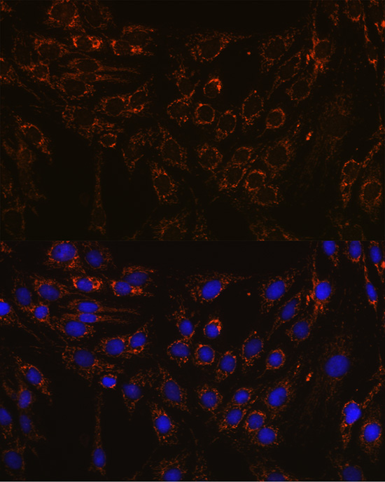
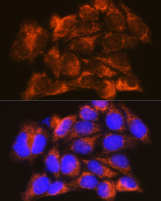
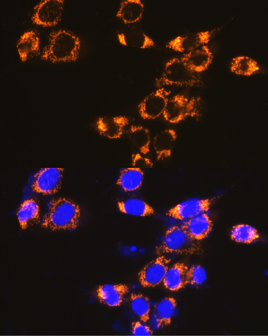
Immunofluorescence analysis of C6 cells using IFNAR1 Rabbit pAb (A18594) at dilution of 1:100 (40x lens). Secondary antibody: Cy3-conjugated Goat anti-Rabbit IgG (H+L) (AS007) at 1:500 dilution. Blue: DAPI for nuclear staining. |
|
|
Immunofluorescence analysis of HeLa cells using IFNAR1 Rabbit pAb (A18594) at dilution of 1:100 (40x lens). Secondary antibody: Cy3-conjugated Goat anti-Rabbit IgG (H+L) (AS007) at 1:500 dilution. Blue: DAPI for nuclear staining. |
|
|
Immunofluorescence analysis of NIH-3T3 cells using IFNAR1 Rabbit pAb (A18594) at dilution of 1:100 (40x lens). Secondary antibody: Cy3-conjugated Goat anti-Rabbit IgG (H+L) (AS007) at 1:500 dilution. Blue: DAPI for nuclear staining. |
Product Guarantee and Expert Support
