
MAP3K1 Rabbit pAb, Unconjugated, Polyclonal
Catalog Number:
ABB-A16057
- Images (8)


| Article Name: | MAP3K1 Rabbit pAb, Unconjugated, Polyclonal |
| Biozol Catalog Number: | ABB-A16057 |
| Supplier Catalog Number: | A16057 |
| Alternative Catalog Number: | ABB-A16057-1000UL,ABB-A16057-500UL,ABB-A16057-100UL,ABB-A16057-20UL |
| Manufacturer: | ABclonal |
| Host: | Rabbit |
| Category: | Antikörper |
| Application: | ELISA, IF, IHC-P, WB |
| Species Reactivity: | Human |
| Immunogen: | Synthetic peptide. This information is considered to be commercially sensitive. |
| Conjugation: | Unconjugated |
| Alternative Names: | MEKK, MEKK1, SRXY6, MEKK 1, MAPKKK1, MAP3K1 |
| The protein encoded by this gene is a serine/threonine kinase and is part of some signal transduction cascades, including the ERK and JNK kinase pathways as well as the NF-kappa-B pathway. The encoded protein is activated by autophosphorylation and requires magnesium as a cofactor in phosphorylating other proteins. This protein has E3 ligase activity conferred by a plant homeodomain (PHD) in its N-terminus and phospho-kinase activity conferred by a kinase domain in its C-terminus. |
| Application Dilute: | WB,1:1000 - 1:4000|IF/ICC,1:50 - 1:100|IF-P,1:50 - 1:100|IHC-P,1:50 - 1:100|ELISA,Recommended starting concentration is 1 µg/mL. Please optimize the concentration based on your specific assay requirements. |
| Application Notes: | Cross-Reactivity: Human,Mouse,Rat. ResearchArea: Cancer,Signal Transduction,Kinase,Serine threonine kinases,ErbB-HER Signaling Pathway,MAPK-JNK Signaling Pathway,Cell Biology Developmental Biology,Cytoskeleton,Actins,Ubiquitin,Ubiquitin-Proteasome Signaling Pathway,TGF-b-Smad Signaling Pathway,Immunology Inflammation,B Cell Receptor Signaling Pathway,T Cell Receptor Signaling Pathway,Toll-like Receptor Signaling Pathway. Shipping: Ice Bag |
|
|
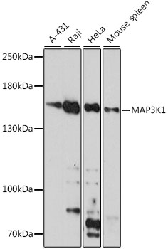
Western blot analysis of various lysates using MAP3K1 Rabbit pAb (A16057) at 1:1000 dilution. Secondary antibody: HRP-conjugated Goat anti-Rabbit IgG (H+L) (AS014) at 1:10000 dilution. Lysates/proteins: 25µg per lane. Blocking buffer: 3% nonfat dry milk in TBST. Detection: ECL Basic Kit (RM00020). Exposure time: 30s. |
|
|
Western blot analysis of various lysates using MAP3K1 Rabbit pAb (A16057) at 1:1000 dilution incubated overnight at 4°C. Secondary antibody: HRP-conjugated Goat anti-Rabbit IgG (H+L) (AS014) at 1:10000 dilution. Lysates/proteins: 25 µg per lane. Blocking buffer: 3% nonfat dry milk in TBST. Detection: ECL Basic Kit (RM00020). Exposure time: 20s. |
|
|
Immunohistochemistry analysis of paraffin-embedded Human pancreas tissue using MAP3K1 Rabbit pAb (A16057) at a dilution of 1:300 (40x lens). High pressure antigen retrieval performed with 0.01M Citrate Buffer (pH 6.0) prior to IHC staining. |
|
|
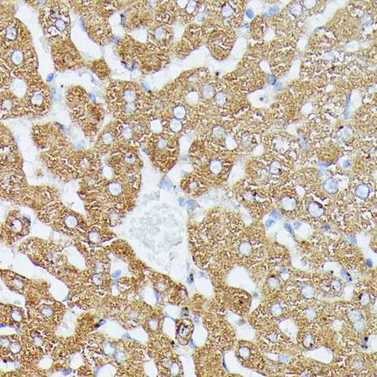
Immunohistochemistry analysis of paraffin-embedded Human liver tissue using MAP3K1 Rabbit pAb (A16057) at a dilution of 1:300 (40x lens). High pressure antigen retrieval performed with 0.01M Citrate Buffer (pH 6.0) prior to IHC staining. |
|
|
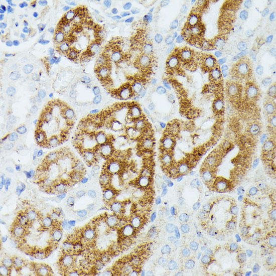
Immunohistochemistry analysis of paraffin-embedded Mouse kidney tissue using MAP3K1 Rabbit pAb (A16057) at a dilution of 1:300 (40x lens). High pressure antigen retrieval performed with 0.01M Citrate Buffer (pH 6.0) prior to IHC staining. |
|
|
Immunohistochemistry analysis of paraffin-embedded Rat kidney tissue using MAP3K1 Rabbit pAb (A16057) at a dilution of 1:300 (40x lens). High pressure antigen retrieval performed with 0.01M Citrate Buffer (pH 6.0) prior to IHC staining. |
|
|
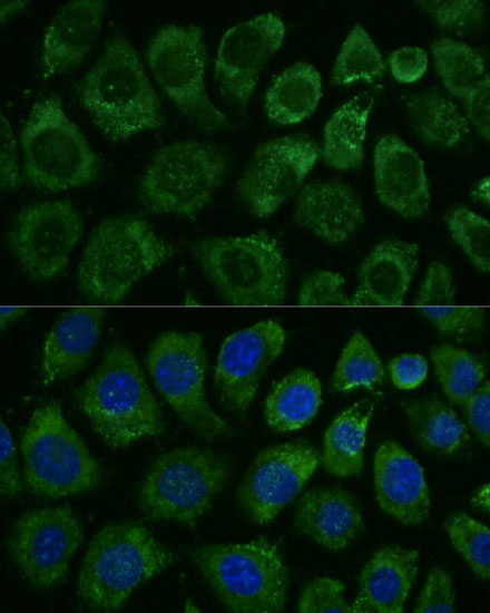
Immunofluorescence analysis of SH-SY5Y cells using MAP3K1 Rabbit pAb (A16057) at a dilution of 1:200 (40x lens). Secondary antibody: Cy3-conjugated Goat anti-Rabbit IgG (H+L)(AS007) at 1:500 dilution. Blue: DAPI for nuclear staining. |
|
|
Immunofluorescence analysis of Mouse spleen tissue using MAP3K1 Rabbit pAb (A16057) at a dilution of 1:200 (40x lens). Secondary antibody: Cy3-conjugated Goat anti-Rabbit IgG (H+L)(AS007) at 1:500 dilution. Blue: DAPI for nuclear staining. High pressure antigen retrieval performed with 0.01M Citrate Buffer(pH 6.0) prior to IF staining. |
Product Guarantee and Expert Support
