
ATP5C1 Rabbit pAb, Unconjugated, Polyclonal
Catalog Number:
ABB-A15257
- Images (7)


| Article Name: | ATP5C1 Rabbit pAb, Unconjugated, Polyclonal |
| Biozol Catalog Number: | ABB-A15257 |
| Supplier Catalog Number: | A15257 |
| Alternative Catalog Number: | ABB-A15257-100UL,ABB-A15257-20UL,ABB-A15257-500UL,ABB-A15257-1000UL |
| Manufacturer: | ABclonal |
| Host: | Rabbit |
| Category: | Antikörper |
| Application: | ELISA, IF, IHC-P, WB |
| Species Reactivity: | Human |
| Immunogen: | Recombinant protein (or fragment).This information is considered to be commercially sensitive. |
| Conjugation: | Unconjugated |
| Alternative Names: | ATP5C, ATP5C1, ATP5CL1 |
| This gene encodes a subunit of mitochondrial ATP synthase. Mitochondrial ATP synthase catalyzes ATP synthesis, utilizing an electrochemical gradient of protons across the inner membrane during oxidative phosphorylation. ATP synthase is composed of two linked multi-subunit complexes: the soluble catalytic core, F1, and the membrane-spanning component, Fo, comprising the proton channel. The catalytic portion of mitochondrial ATP synthase consists of 5 different subunits (alpha, beta, gamma, delta, and epsilon) assembled with a stoichiometry of 3 alpha, 3 beta, and a single representative of the other 3. The proton channel consists of three main subunits (a, b, c). This gene encodes the gamma subunit of the catalytic core. Alternatively spliced transcript variants encoding different isoforms have been identified. This gene also has a pseudogene on chromosome 14. |
| Clonality: | Polyclonal |
| Molecular Weight: | 33kDa |
| NCBI: | 509 |
| UniProt: | P36542 |
| Purity: | Affinity purification |
| Sequence: | ATLKDITRRLKSIKNIQKITKSMKMVAAAKYARAERELKPARIYGLGSLALYEKADIKGPEDKKKHLLIGVSSDRGLCGAIHSSIAKQMKSEVATLTAAGKEVMLVGIGDKIRGILYRTHSDQFLVAFKEVGRKPPTFGDASVIALELLNSGYEFDEGSIIFNKFRSVISYKTEEKPIFSLNTVASADSMSIYDDIDADVLQNYQEYNLANIIYYSLKESTTSEQSARMTAMDNASKNASEMIDKLTLTFNRTRQ |
| Target: | ATP5F1C |
| Antibody Type: | Primary Antibody |
| Application Dilute: | WB,1:200 - 1:2000|IHC-P,1:50 - 1:200|IF/ICC,1:50 - 1:200|ELISA,Recommended starting concentration is 1 µg/mL. Please optimize the concentration based on your specific assay requirements. |
| Application Notes: | Cross-Reactivity: Human,Mouse,Rat. ResearchArea: Epigenetics Nuclear Signaling,RNA Binding,Signal Transduction,Endocrine Metabolism,Mitochondrial metabolism,Mitochondrial markers,Neuroscience,Neurodegenerative Diseases. Shipping: Ice Bag |
|
|
Immunohistochemistry analysis of paraffin-embedded Rat ovary using ATP5C1 Rabbit pAb (A15257) at dilution of 1:100 (40x lens). Microwave antigen retrieval performed with 0.01M PBS Buffer (pH 7.2) prior to IHC staining. |
|
|
Immunohistochemistry analysis of paraffin-embedded Human lung cancer using ATP5C1 Rabbit pAb (A15257) at dilution of 1:100 (40x lens). Microwave antigen retrieval performed with 0.01M PBS Buffer (pH 7.2) prior to IHC staining. |
|
|
Western blot analysis of various lysates, using ATP5C1 Rabbit pAb (A15257) at 1:1000 dilution. Secondary antibody: HRP-conjugated Goat anti-Rabbit IgG (H+L) (AS014) at 1:10000 dilution. Lysates/proteins: 25µg per lane. Blocking buffer: 3% nonfat dry milk in TBST. Detection: ECL Basic Kit (RM00020). Exposure time: 90s. |
|
|
Immunohistochemistry analysis of paraffin-embedded Human placenta using ATP5C1 Rabbit pAb (A15257) at dilution of 1:100 (40x lens). Microwave antigen retrieval performed with 0.01M PBS Buffer (pH 7.2) prior to IHC staining. |
|
|
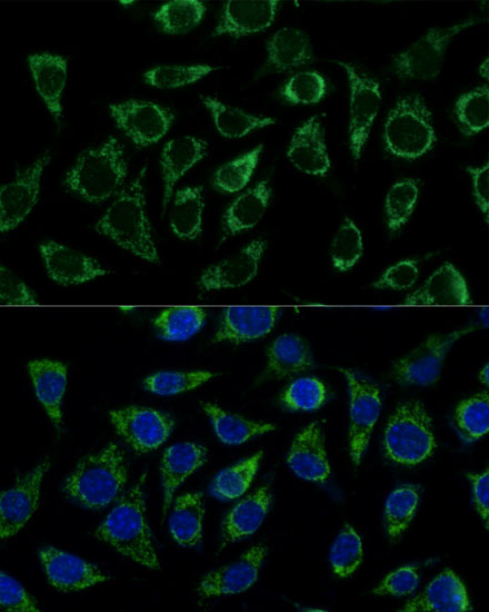
Immunofluorescence analysis of C6 cells using ATP5C1 Rabbit pAb (A15257) at dilution of 1:100 (40x lens). Secondary antibody: Cy3-conjugated Goat anti-Rabbit IgG (H+L) (AS007) at 1:500 dilution. Blue: DAPI for nuclear staining. |
|
|
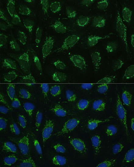
Immunofluorescence analysis of L929 cells using ATP5C1 Rabbit pAb (A15257) at dilution of 1:100 (40x lens). Secondary antibody: Cy3-conjugated Goat anti-Rabbit IgG (H+L) (AS007) at 1:500 dilution. Blue: DAPI for nuclear staining. |
|
|
Immunofluorescence analysis of U-2 OS cells using ATP5C1 Rabbit pAb (A15257) at dilution of 1:100 (40x lens). Secondary antibody: Cy3-conjugated Goat anti-Rabbit IgG (H+L) (AS007) at 1:500 dilution. Blue: DAPI for nuclear staining. |
Product Guarantee and Expert Support
