
Ataxin-3 (ATXN3) Rabbit pAb, Unconjugated, Polyclonal
Catalog Number:
ABB-A1243
- Images (7)


| Article Name: | Ataxin-3 (ATXN3) Rabbit pAb, Unconjugated, Polyclonal |
| Biozol Catalog Number: | ABB-A1243 |
| Supplier Catalog Number: | A1243 |
| Alternative Catalog Number: | ABB-A1243-20UL,ABB-A1243-100UL,ABB-A1243-500UL,ABB-A1243-1000UL |
| Manufacturer: | ABclonal |
| Host: | Rabbit |
| Category: | Antikörper |
| Application: | ELISA, IF, IHC-P, WB |
| Species Reactivity: | Human |
| Immunogen: | Recombinant protein (or fragment).This information is considered to be commercially sensitive. |
| Conjugation: | Unconjugated |
| Alternative Names: | AT3, JOS, MJD, ATX3, MJD1, SCA3, Ataxin-3 (ATXN3) |
| Machado-Joseph disease, also known as spinocerebellar ataxia-3, is an autosomal dominant neurologic disorder. The protein encoded by this gene contains (CAG)n repeats in the coding region, and the expansion of these repeats from the normal 12-44 to 52-86 is one cause of Machado-Joseph disease. There is a negative correlation between the age of onset and CAG repeat numbers. Alternatively spliced transcript variants encoding different isoforms have been described for this gene. |
| Clonality: | Polyclonal |
| Molecular Weight: | 41kDa |
| NCBI: | 4287 |
| UniProt: | P54252 |
| Purity: | Affinity purification |
| Sequence: | MESIFHEKQEGSLCAQHCLNNLLQGEYFSPVELSSIAHQLDEEERMRMAEGGVTSEDYRTFLQQPSGNMDDSGFFSIQVISNALKVWGLELILFNSPEYQRLRIDPINERSFICNYKEHWFTVRKLGKQWFNLNSLLTGPELISDTYLALFLAQLQQEGYSIFVVKGDLPDCEADQLLQMIRVQQMHRPKLIGEELAQLKEQRVHKTDLERVLEANDGSGMLDEDEEDLQRALALSRQEIDMEDEEADLRRAIQL |
| Target: | ATXN3 |
| Antibody Type: | Primary Antibody |
| Application Dilute: | WB,1:500 - 1:1000|IHC-P,1:50 - 1:200|IF/ICC,1:50 - 1:200|ELISA,Recommended starting concentration is 1 µg/mL. Please optimize the concentration based on your specific assay requirements. |
| Application Notes: | Cross-Reactivity: Human,Mouse,Rat. ResearchArea: Epigenetics Nuclear Signaling,Cell Biology Developmental Biology,Ubiquitin,Neuroscience,Neurodegenerative Diseases. Shipping: Ice Bag |
|
|
Immunohistochemistry analysis of paraffin-embedded Human esophageal cancer using Ataxin-3 (ATXN3) Rabbit pAb (A1243) at dilution of 1:100 (40x lens). High pressure antigen retrieval performed with 0.01M Citrate buffer (pH 6.0) prior to IHC staining. |
|
|
Immunohistochemistry analysis of paraffin-embedded Mouse lung using Ataxin-3 (ATXN3) Rabbit pAb (A1243) at dilution of 1:100 (40x lens). High pressure antigen retrieval performed with 0.01M Citrate buffer (pH 6.0) prior to IHC staining. |
|
|
Western blot analysis of various lysates using Ataxin-3 (ATXN3) Rabbit pAb (A1243) at 1:1000 dilution. Secondary antibody: HRP-conjugated Goat anti-Rabbit IgG (H+L) (AS014) at 1:10000 dilution. Lysates/proteins: 25µg per lane. Blocking buffer: 3% nonfat dry milk in TBST. Detection: ECL Basic Kit (RM00020). Exposure time: 10s. |
|
|
Immunohistochemistry analysis of paraffin-embedded Rat kidney using Ataxin-3 (ATXN3) Rabbit pAb (A1243) at dilution of 1:100 (40x lens). High pressure antigen retrieval performed with 0.01M Citrate buffer (pH 6.0) prior to IHC staining. |
|
|
Confocal immunofluorescence analysis of U-2 OS cells using Ataxin-3 (ATXN3) Rabbit pAb (A1243) at dilution of 1:100. Blue: DAPI for nuclear staining. |
|
|
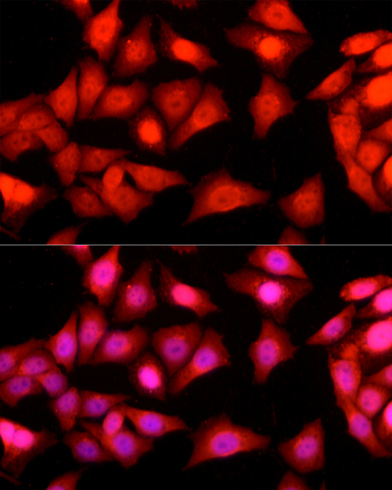
Immunofluorescence analysis of HeLa cells using Ataxin-3 (ATXN3) Rabbit pAb (A1243) at dilution of 1:100 (40x lens). Secondary antibody: Cy3-conjugated Goat anti-Rabbit IgG (H+L) (AS007) at 1:500 dilution. Blue: DAPI for nuclear staining. |
|
|
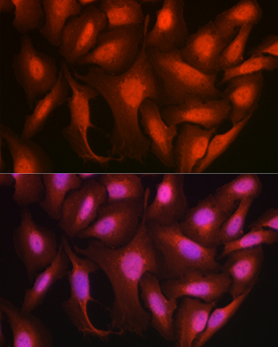
Immunofluorescence analysis of U2OS cells using Ataxin-3 (ATXN3) Rabbit pAb (A1243) at dilution of 1:100 (40x lens). Secondary antibody: Cy3-conjugated Goat anti-Rabbit IgG (H+L) (AS007) at 1:500 dilution. Blue: DAPI for nuclear staining. |
Product Guarantee and Expert Support
