
TAK1 Rabbit pAb, Unconjugated, Polyclonal
Catalog Number:
ABB-A12022
- Images (7)


| Article Name: | TAK1 Rabbit pAb, Unconjugated, Polyclonal |
| Biozol Catalog Number: | ABB-A12022 |
| Supplier Catalog Number: | A12022 |
| Alternative Catalog Number: | ABB-A12022-100UL,ABB-A12022-20UL,ABB-A12022-1000UL,ABB-A12022-500UL |
| Manufacturer: | ABclonal |
| Host: | Rabbit |
| Category: | Antikörper |
| Application: | ELISA, IF, IHC-P, WB |
| Species Reactivity: | Human |
| Immunogen: | Synthetic peptide. This information is considered to be commercially sensitive. |
| Conjugation: | Unconjugated |
| Alternative Names: | CSCF, FMD2, TAK1, MEKK7, TGF1a |
| The protein encoded by this gene is a member of the serine/threonine protein kinase family. This kinase mediates the signaling transduction induced by TGF beta and morphogenetic protein (BMP), and controls a variety of cell functions including transcription regulation and apoptosis. In response to IL-1, this protein forms a kinase complex including TRAF6, MAP3K7P1/TAB1 and MAP3K7P2/TAB2, this complex is required for the activation of nuclear factor kappa B. This kinase can also activate MAPK8/JNK, MAP2K4/MKK4, and thus plays a role in the cell response to environmental stresses. Four alternatively spliced transcript variants encoding distinct isoforms have been reported. |
| Application Dilute: | WB,1:500 - 1:1000|IHC-P,1:50 - 1:200|IF/ICC,1:50 - 1:200|ELISA,Recommended starting concentration is 1 µg/mL. Please optimize the concentration based on your specific assay requirements. |
| Application Notes: | Cross-Reactivity: Human,Mouse,Rat. ResearchArea: Protein phosphorylation,Cancer,Signal Transduction,G protein signaling,G-Protein-Coupled Receptors Signaling to MAPK Erk,Kinase,Serine threonine kinases,Tyrosine kinases,MAPK-Erk Signaling Pathway,MAPK-JNK Signaling Pathway,Cell Biology Developmental Biology,Apoptosis,Inhibition of Apoptosis,Cytoskeleton,Actins,TGF-b-Smad Signaling Pathway,Wnt -Catenin Signaling Pathway,Immunology Inflammation,B Cell Receptor Signaling Pathway,T Cell Receptor Signaling Pathway,NF-kB Signaling Pathway,Toll-like Receptor Signaling Pathway,Cell Intrinsic Innate Immunity Signaling Pathway,TLR Signaling,Cardiovascular. Shipping: Ice Bag |
|
|
Western blot analysis of lysates from Mouse brain usingTAK1 Rabbit pAb (A12022) at1:1000 dilution. Secondary antibody: HRP-conjugated Goat anti-Rabbit IgG (H+L) (AS014) at 1:10000 dilution. Lysates/proteins: 25µg per lane. Blocking buffer: 3% nonfat dry milk in TBST. Detection: ECL Basic Kit (RM00020). Exposure time:10s. |
|
|
Immunohistochemistry analysis of paraffin-embedded Human colon carcinoma using TAK1 Rabbit pAb (A12022) at dilution of 1:50 (40x lens). High pressure antigen retrieval performed with 0.01M Citrate buffer (pH 6.0) prior to IHC staining. |
|
|
Western blot analysis of various lysates, using TAK1 Rabbit pAb (A12022) at 1:400 dilution. Secondary antibody: HRP-conjugated Goat anti-Rabbit IgG (H+L) (AS014) at 1:10000 dilution. Lysates/proteins: 25µg per lane. Blocking buffer: 3% nonfat dry milk in TBST. Detection: ECL Basic Kit (RM00020). Exposure time: 30s. |
|
|
Immunohistochemistry analysis of paraffin-embedded Mouse kidney using TAK1 Rabbit pAb (A12022) at dilution of 1:50 (40x lens). High pressure antigen retrieval performed with 0.01M Citrate buffer (pH 6.0) prior to IHC staining. |
|
|
Immunofluorescence analysis of NIH/3T3 cells using TAK1 Rabbit pAb (A12022) at dilution of 1:50 (40x lens). Secondary antibody: Cy3-conjugated Goat anti-Rabbit IgG (H+L) (AS007) at 1:500 dilution. Blue: DAPI for nuclear staining. |
|
|
Immunofluorescence analysis of PC-12 cells using TAK1 Rabbit pAb (A12022) at dilution of 1:50 (40x lens). Secondary antibody: Cy3-conjugated Goat anti-Rabbit IgG (H+L) (AS007) at 1:500 dilution. Blue: DAPI for nuclear staining. |
|
|
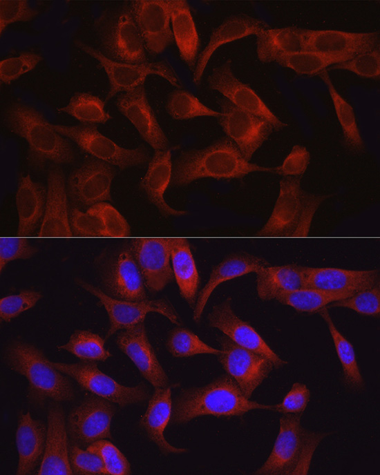
Immunofluorescence analysis of U2OS cells using TAK1 Rabbit pAb (A12022) at dilution of 1:50 (40x lens). Secondary antibody: Cy3-conjugated Goat anti-Rabbit IgG (H+L) (AS007) at 1:500 dilution. Blue: DAPI for nuclear staining. |
Product Guarantee and Expert Support
