
TLR3 Rabbit pAb, Unconjugated, Polyclonal
Catalog Number:
ABB-A11778
- Images (7)


| Article Name: | TLR3 Rabbit pAb, Unconjugated, Polyclonal |
| Biozol Catalog Number: | ABB-A11778 |
| Supplier Catalog Number: | A11778 |
| Alternative Catalog Number: | ABB-A11778-100UL,ABB-A11778-20UL,ABB-A11778-500UL,ABB-A11778-1000UL |
| Manufacturer: | ABclonal |
| Host: | Rabbit |
| Category: | Antikörper |
| Application: | ELISA, IF, IHC-P, WB |
| Species Reactivity: | Human |
| Immunogen: | Recombinant protein (or fragment).This information is considered to be commercially sensitive. |
| Conjugation: | Unconjugated |
| Alternative Names: | CD283, IIAE2, IMD83, TLR3 |
| The protein encoded by this gene is a member of the Toll-like receptor (TLR) family which plays a fundamental role in pathogen recognition and activation of innate immunity. TLRs are highly conserved from Drosophila to humans and share structural and functional similarities. They recognize pathogen-associated molecular patterns (PAMPs) that are expressed on infectious agents, and mediate the production of cytokines necessary for the development of effective immunity. The various TLRs exhibit different patterns of expression. This receptor is most abundantly expressed in placenta and pancreas, and is restricted to the dendritic subpopulation of the leukocytes. It recognizes dsRNA associated with viral infection, and induces the activation of NF-kappaB and the production of type I interferons. It thus plays a role in host defense against multiple viruses. |
| Clonality: | Polyclonal |
| Molecular Weight: | 104kDa |
| NCBI: | 7098 |
| UniProt: | O15455 |
| Purity: | Affinity purification |
| Sequence: | SPFQPLRNLTILDLSNNNIANINDDMLEGLEKLEILDLQHNNLARLWKHANPGGPIYFLKGLSHLHILNLESNGFDEIPVEVFKDLFELKIIDLGLNNLNTLPASVFNNQVSLKSLNLQKNLITSVEKKVFGPAFRNLTELDMRFNPFDCTCESIAWFVNWINETHTNIPELSSHYLCNTPPHYHGFPVRLFDTSSCKDSA |
| Target: | TLR3 |
| Antibody Type: | Primary Antibody |
| Application Dilute: | WB,1:500 - 1:1000|IHC-P,1:50 - 1:200|IF/ICC,1:50 - 1:200|ELISA,Recommended starting concentration is 1 µg/mL. Please optimize the concentration based on your specific assay requirements. |
| Application Notes: | Cross-Reactivity: Human,Mouse,Rat. ResearchArea: Signal Transduction,Immunology Inflammation,CDs,NF-kB Signaling Pathway,Toll-like Receptor Signaling Pathway,Cell Intrinsic Innate Immunity Signaling Pathway,TLR Signaling. Shipping: Ice Bag |
|
|
Western blot analysis of various lysates using TLR3 Rabbit pAb (A11778) at 1:500 dilution. Secondary antibody: HRP-conjugated Goat anti-Rabbit IgG (H+L) (AS014) at 1:10000 dilution. Lysates/proteins: 25µg per lane. Blocking buffer: 3% nonfat dry milk in TBST. Detection: ECL Basic Kit (RM00020). Exposure time: 10s. |
|
|
Western blot analysis of lysates from Mouse lung, using TLR3 Rabbit pAb (A11778) at 1:500 dilution. Secondary antibody: HRP-conjugated Goat anti-Rabbit IgG (H+L) (AS014) at 1:10000 dilution. Lysates/proteins: 25µg per lane. Blocking buffer: 3% nonfat dry milk in TBST. Detection: ECL Basic Kit (RM00020). Exposure time: 180s. |
|
|
Western blot analysis of various lysates using TLR3 Rabbit pAb (A11778) at 1:1000 dilution. Secondary antibody: HRP-conjugated Goat anti-Rabbit IgG (H+L) (AS014) at 1:10000 dilution. Lysates/proteins: 25µg per lane. Blocking buffer: 3% nonfat dry milk in TBST. Detection: ECL Basic Kit (RM00020). Exposure time: 90s. |
|
|
Immunohistochemistry analysis of paraffin-embedded Human placenta using TLR3 Rabbit pAb (A11778) at dilution of 1:100 (40x lens). Microwave antigen retrieval performed with 0.01M Tris/EDTA Buffer (pH 9.0) prior to IHC staining. |
|
|
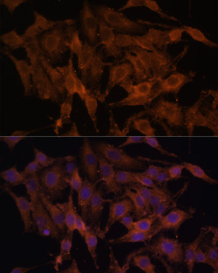
Immunofluorescence analysis of C6 cells using TLR3 Rabbit pAb (A11778) at dilution of 1:100. Secondary antibody: Cy3-conjugated Goat anti-Rabbit IgG (H+L) (AS007) at 1:500 dilution. Blue: DAPI for nuclear staining. |
|
|
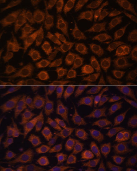
Immunofluorescence analysis of HeLa cells using TLR3 Rabbit pAb (A11778) at dilution of 1:100. Secondary antibody: Cy3-conjugated Goat anti-Rabbit IgG (H+L) (AS007) at 1:500 dilution. Blue: DAPI for nuclear staining. |
|
|
Immunofluorescence analysis of L929 cells using TLR3 Rabbit pAb (A11778) at dilution of 1:100. Secondary antibody: Cy3-conjugated Goat anti-Rabbit IgG (H+L) (AS007) at 1:500 dilution. Blue: DAPI for nuclear staining. |
Product Guarantee and Expert Support
