
hnRNP A1 Rabbit mAb, Unconjugated, Monoclonal
Catalog Number:
ABB-A11564
- Images (7)


| Article Name: | hnRNP A1 Rabbit mAb, Unconjugated, Monoclonal |
| Biozol Catalog Number: | ABB-A11564 |
| Supplier Catalog Number: | A11564 |
| Alternative Catalog Number: | ABB-A11564-100UL,ABB-A11564-20UL,ABB-A11564-1000UL,ABB-A11564-500UL |
| Manufacturer: | ABclonal |
| Host: | Rabbit |
| Category: | Antikörper |
| Application: | ELISA, IF, IHC-P, IP, WB |
| Species Reactivity: | Human |
| Immunogen: | Synthetic peptide. This information is considered to be commercially sensitive. |
| Conjugation: | Unconjugated |
| Alternative Names: | UP 1, ALS19, ALS20, HNRPA1, IBMPFD3, HNRPA1L3, hnRNP A1, hnRNP-A1 |
| This gene encodes a member of a family of ubiquitously expressed heterogeneous nuclear ribonucleoproteins (hnRNPs), which are RNA-binding proteins that associate with pre-mRNAs in the nucleus and influence pre-mRNA processing, as well as other aspects of mRNA metabolism and transport. The protein encoded by this gene is one of the most abundant core proteins of hnRNP complexes and plays a key role in the regulation of alternative splicing. Mutations in this gene have been observed in individuals with amyotrophic lateral sclerosis 20. Multiple alternatively spliced transcript variants have been found. There are numerous pseudogenes of this gene distributed throughout the genome.hnRNP A1 has three isoforms with MW 30 kDa, 34 kDa and 39 kDa. |
| Application Dilute: | WB,1:1000 - 1:2000|IHC-P,1:100 - 1:1000|IF/ICC,1:200 - 1:2000|IP,0.5µg-4µg antibody for 200µg-400µg extracts of whole cells|ELISA,Recommended starting concentration is 1 µg/mL. Please optimize the concentration based on your specific assay requirements. |
| Application Notes: | Cross-Reactivity: Human,Mouse,Rat. ResearchArea: Epigenetics Nuclear Signaling,RNA Binding. Shipping: Ice Bag |
|
|
Western blot analysis of various lysates using hnRNP A1 Rabbit mAb (A11564) at 1:1000 dilution. Secondary antibody: HRP-conjugated Goat anti-Rabbit IgG (H+L) (AS014) at 1:10000 dilution. Lysates/proteins: 25µg per lane. Blocking buffer: 3% nonfat dry milk in TBST. Detection: ECL Basic Kit (RM00020). Exposure time: 3min. |
|
|
Immunohistochemistry analysis of paraffin-embedded Human colon using hnRNP A1 Rabbit mAb (A11564) at dilution of 1:100 (40x lens). Microwave antigen retrieval performed with 0.01M PBS Buffer (pH 7.2) prior to IHC staining. |
|
|
Western blot analysis of various lysates using hnRNP A1 Rabbit mAb (A11564) at 1:1000 dilution. Secondary antibody: HRP-conjugated Goat anti-Rabbit IgG (H+L) (AS014) at 1:10000 dilution. Lysates/proteins: 25µg per lane. Blocking buffer: 3% nonfat dry milk in TBST. Detection: ECL Basic Kit (RM00020). Exposure time: 60s. |
|
|
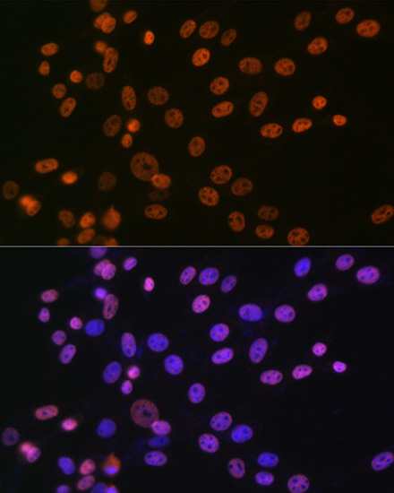
Confocal imaging of U-2 OS cells usinghnRNP A1 Rabbit mAb (A11564,dilution 1:200) followed by a further incubation with Cy3 Goat Anti-Rabbit IgG (H+L) (AS007,dilution 1:500)(Red).The cells were counterstained with alpha-Tubulin Mouse mAb (AC012, dilution 1:400) followed by incubation with ABflo 488-conjugated Goat Anti-Mouse IgG (H+L) Ab (AS076, dilution 1:500) (Green).DAPI was used for nuclear staining (Blue). Objective: 100x. |
|
|
Confocal imaging of NIH/3T3 cells usinghnRNP A1 Rabbit mAb (A11564,dilution 1:200) followed by a further incubation with Cy3 Goat Anti-Rabbit IgG (H+L) (AS007,dilution 1:500)(Red).The cells were counterstained with alpha-Tubulin Mouse mAb (AC012, dilution 1:400) followed by incubation with ABflo 488-conjugated Goat Anti-Mouse IgG (H+L) Ab (AS076, dilution 1:500) (Green).DAPI was used for nuclear staining (Blue). Objective: 100x. |
|
|
Confocal imaging of C6 cells usinghnRNP A1 Rabbit mAb (A11564,dilution 1:200) followed by a further incubation with Cy3 Goat Anti-Rabbit IgG (H+L) (AS007,dilution 1:500)(Red).The cells were counterstained with alpha-Tubulin Mouse mAb (AC012, dilution 1:400) followed by incubation with ABflo 488-conjugated Goat Anti-Mouse IgG (H+L) Ab (AS076, dilution 1:500) (Green).DAPI was used for nuclear staining (Blue). Objective: 100x. |
|
|
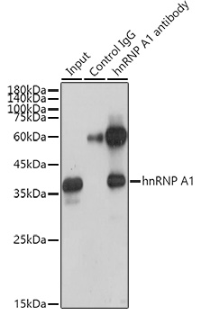
Immunoprecipitation analysis of 300 µg extracts of HeLa cells using 3 µg hnRNP A1 antibody (A11564). Western blot was performed from the immunoprecipitate using hnRNP A1 antibody (A11564) at a dilution of 1:1000. |
Product Guarantee and Expert Support
