
GC1q R/C1QBP Rabbit mAb, Unconjugated, Monoclonal
Catalog Number:
ABB-A11292
- Images (7)


| Article Name: | GC1q R/C1QBP Rabbit mAb, Unconjugated, Monoclonal |
| Biozol Catalog Number: | ABB-A11292 |
| Supplier Catalog Number: | A11292 |
| Alternative Catalog Number: | ABB-A11292-100UL,ABB-A11292-20UL |
| Manufacturer: | ABclonal |
| Host: | Rabbit |
| Category: | Antikörper |
| Application: | ELISA, IHC-P, WB |
| Species Reactivity: | Human |
| Immunogen: | Synthetic peptide. This information is considered to be commercially sensitive. |
| Conjugation: | Unconjugated |
| Alternative Names: | p32, HABP1, gC1qR, GC1QBP, SF2p32, gC1Q-R, COXPD33, SF2AP32, GC1q R/C1QBP |
| The human complement subcomponent C1q associates with C1r and C1s in order to yield the first component of the serum complement system. The protein encoded by this gene is known to bind to the globular heads of C1q molecules and inhibit C1 activation. This protein has also been identified as the p32 subunit of pre-mRNA splicing factor SF2, as well as a hyaluronic acid-binding protein. |
| Application Dilute: | WB,1:500 - 1:1000|IHC-P,1:50 - 1:200|ELISA,Recommended starting concentration is 1 µg/mL. Please optimize the concentration based on your specific assay requirements. |
| Application Notes: | Cross-Reactivity: Human,Mouse,Rat. ResearchArea: Immunology Inflammation,Cell Intrinsic Innate Immunity Signaling Pathway. Shipping: Ice Bag |
|
|
Western blot analysis of various lysates using GC1q R/C1QBP Rabbit mAb (A11292) at 1:1000 dilution. Secondary antibody: HRP-conjugated Goat anti-Rabbit IgG (H+L) (AS014) at 1:10000 dilution. Lysates/proteins: 25µg per lane. Blocking buffer: 3% nonfat dry milk in TBST. Detection: ECL Basic Kit (RM00020). Exposure time: 10s. |
|
|
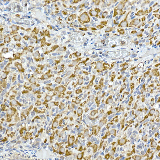
Immunohistochemistry analysis of paraffin-embeddedHuman thyroid cancer tissue usingGC1q R/C1QBP Rabbit mAb(A11292) at a dilution of 1:200 (40x lens).High pressure antigen retrieval was performed with 0.01 M citrate buffer (pH 6.0) prior to IHC staining. |
|
|
Immunohistochemistry analysis of paraffin-embeddedHuman liver tissue usingGC1q R/C1QBP Rabbit mAb(A11292) at a dilution of 1:200 (40x lens).High pressure antigen retrieval was performed with 0.01 M citrate buffer (pH 6.0) prior to IHC staining. |
|
|
Immunohistochemistry analysis of paraffin-embeddedHuman colon carcinoma tissue usingGC1q R/C1QBP Rabbit mAb(A11292) at a dilution of 1:200 (40x lens).High pressure antigen retrieval was performed with 0.01 M citrate buffer (pH 6.0) prior to IHC staining. |
|
|
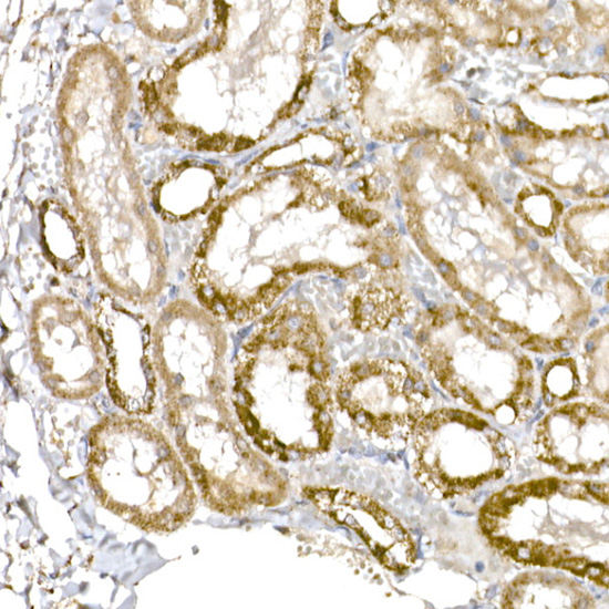
Immunohistochemistry analysis of paraffin-embeddedRat kidney tissue usingGC1q R/C1QBP Rabbit mAb(A11292) at a dilution of 1:200 (40x lens).High pressure antigen retrieval was performed with 0.01 M citrate buffer (pH 6.0) prior to IHC staining. |
|
|
Immunohistochemistry analysis of paraffin-embeddedHuman colon tissue usingGC1q R/C1QBP Rabbit mAb(A11292) at a dilution of 1:200 (40x lens).High pressure antigen retrieval was performed with 0.01 M citrate buffer (pH 6.0) prior to IHC staining. |
|
|
Immunohistochemistry analysis of paraffin-embeddedMouse kidney tissue usingGC1q R/C1QBP Rabbit mAb(A11292) at a dilution of 1:200 (40x lens).High pressure antigen retrieval was performed with 0.01 M citrate buffer (pH 6.0) prior to IHC staining. |
Product Guarantee and Expert Support
