
LC3B Rabbit pAb, Unconjugated, Polyclonal
Catalog Number:
ABB-A11282
- Images (7)

| Article Name: | LC3B Rabbit pAb, Unconjugated, Polyclonal |
| Biozol Catalog Number: | ABB-A11282 |
| Supplier Catalog Number: | A11282 |
| Alternative Catalog Number: | ABB-A11282-20UL,ABB-A11282-100UL,ABB-A11282-1000UL,ABB-A11282-500UL |
| Manufacturer: | ABclonal |
| Host: | Rabbit |
| Category: | Antikörper |
| Application: | ELISA, IF, IHC-P, WB |
| Species Reactivity: | Human |
| Immunogen: | Recombinant protein (or fragment).This information is considered to be commercially sensitive. |
| Conjugation: | Unconjugated |
| Alternative Names: | LC3B, ATG8F, MAP1LC3B-a, MAP1A/1BLC3 |
| The product of this gene is a subunit of neuronal microtubule-associated MAP1A and MAP1B proteins, which are involved in microtubule assembly and important for neurogenesis. Studies on the rat homolog implicate a role for this gene in autophagy, a process that involves the bulk degradation of cytoplasmic component. |
| Application Dilute: | WB,1:500 - 1:1000|IHC-P,1:50 - 1:200|IF/ICC,1:50 - 1:200|ELISA,Recommended starting concentration is 1 µg/mL. Please optimize the concentration based on your specific assay requirements. |
| Application Notes: | Cross-Reactivity: Human,Mouse,Rat. ResearchArea: Cancer,Signal Transduction,Cell Biology Developmental Biology,Autophagy,Cytoskeleton,Microtubules,Endocrine Metabolism,Mitochondrial metabolism,Neuroscience,Cardiovascular,Heart. Shipping: Ice Bag |
|
|
Western blot analysis of lysates from 293T cells usingLC3B Rabbit pAb (A11282) at1:1000 dilution. 293T cells were treated with Chloroquine (50 µM) at 37°C for 20 hours. Secondary antibody: HRP-conjugated Goat anti-Rabbit IgG (H+L) (AS014) at 1:10000 dilution. Lysates/proteins: 25µg per lane. Blocking buffer: 3% nonfat dry milk in TBST. Detection: ECL Basic Kit (RM00020). Exposure time:60s. |
|
|
Western blot analysis of lysates from C6 cells usingLC3B Rabbit pAb (A11282) at1:1000 dilution. C6 cells were treated with Chloroquine (50 µM) at 37°C for 20 hours. Secondary antibody: HRP-conjugated Goat anti-Rabbit IgG (H+L) (AS014) at 1:10000 dilution. Lysates/proteins: 25µg per lane. Blocking buffer: 3% nonfat dry milk in TBST. Detection: ECL Basic Kit (RM00020). Exposure time:60s. |
|
|
Western blot analysis of lysates from NIH/3T3 cells usingLC3B Rabbit pAb (A11282) at1:1000 dilution. NIH/3T3 cells were treated with Chloroquine (50 µM) at 37°C for 20 hours. Secondary antibody: HRP-conjugated Goat anti-Rabbit IgG (H+L) (AS014) at 1:10000 dilution. Lysates/proteins: 25µg per lane. Blocking buffer: 3% nonfat dry milk in TBST. Detection: ECL Basic Kit (RM00020). Exposure time:60s. |
|
|
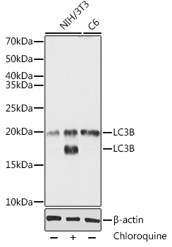
Western blot analysis of various lysates using LC3B Rabbit pAb (A11282)at 1:1000 dilutionincubated overnight at 4°C. 293T,NIH/3T3,C6 cells were treated with Chloroquine at 37°C for 48 hours. Secondary antibody: HRP-conjugated Goat anti-Rabbit IgG (H+L) (AS014) at 1:10000 dilution. Lysates/proteins: 30 µg per lane. Blocking buffer: 3% nonfat dry milk in TBST. Detection: ECL Basic Kit (RM00020). Exposure time: 30s. |
|
|
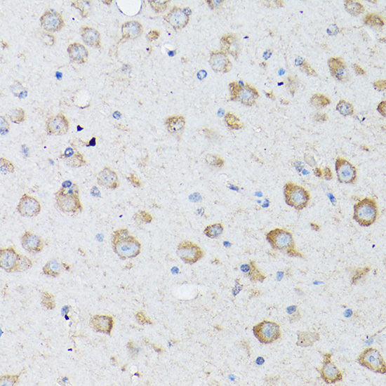
Immunohistochemistry analysis of paraffin-embedded Mouse brain using LC3B Rabbit pAb (A11282) at dilution of 1:100 (40x lens). High pressure antigen retrieval performed with 0.01M Citrate buffer (pH 6.0) prior to IHC staining. |
|
|
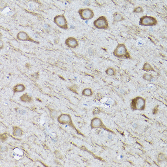
Immunohistochemistry analysis of paraffin-embedded Rat brain using LC3B Rabbit pAb (A11282) at dilution of 1:100 (40x lens). High pressure antigen retrieval performed with 0.01M Citrate buffer (pH 6.0) prior to IHC staining. |
|
|
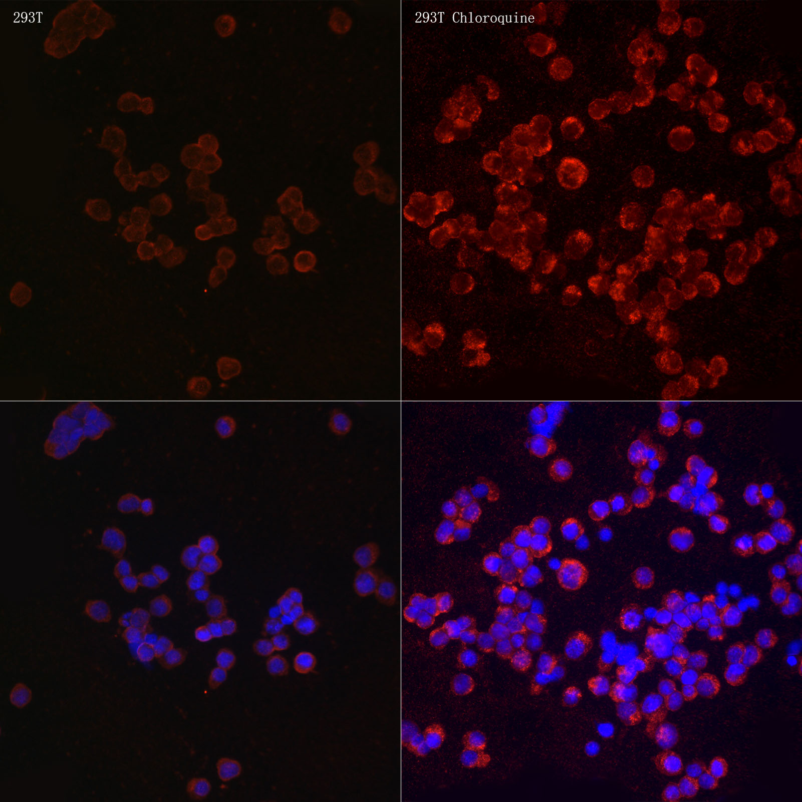
Immunofluorescence analysis of 293T and 293T Chloroquine cells using LC3B Rabbit pAb (A11282) at dilution of 1:100 (40x lens). Secondary antibody: Cy3-conjugated Goat anti-Rabbit IgG (H+L) (AS007) at 1:500 dilution. Blue: DAPI for nuclear staining. |
Product Guarantee and Expert Support
