
SLC27A2 Rabbit pAb, Unconjugated, Polyclonal
Catalog Number:
ABB-A1077
- Images (7)


| Article Name: | SLC27A2 Rabbit pAb, Unconjugated, Polyclonal |
| Biozol Catalog Number: | ABB-A1077 |
| Supplier Catalog Number: | A1077 |
| Alternative Catalog Number: | ABB-A1077-20UL,ABB-A1077-100UL,ABB-A1077-500UL,ABB-A1077-1000UL |
| Manufacturer: | ABclonal |
| Host: | Rabbit |
| Category: | Antikörper |
| Application: | ELISA, IF, IHC-P, WB |
| Species Reactivity: | Human |
| Immunogen: | Recombinant protein (or fragment).This information is considered to be commercially sensitive. |
| Conjugation: | Unconjugated |
| Alternative Names: | VLCS, FATP2, VLACS, ACSVL1, FACVL1, hFACVL1, HsT17226, SLC27A2 |
| The protein encoded by this gene is an isozyme of long-chain fatty-acid-coenzyme A ligase family. Although differing in substrate specificity, subcellular localization, and tissue distribution, all isozymes of this family convert free long-chain fatty acids into fatty acyl-CoA esters, and thereby play a key role in lipid biosynthesis and fatty acid degradation. This isozyme activates long-chain, branched-chain and very-long-chain fatty acids containing 22 or more carbons to their CoA derivatives. It is expressed primarily in liver and kidney, and is present in both endoplasmic reticulum and peroxisomes, but not in mitochondria. Its decreased peroxisomal enzyme activity is in part responsible for the biochemical pathology in X-linked adrenoleukodystrophy. Alternatively spliced transcript variants encoding different isoforms have been found for this gene. |
| Clonality: | Polyclonal |
| Molecular Weight: | 70kDa |
| NCBI: | 11001 |
| UniProt: | O14975 |
| Purity: | Affinity purification |
| Sequence: | TKFSASQFWDDCRKYNVTVIQYIGELLRYLCNSPQKPNDRDHKVRLALGNGLRGDVWRQFVKRFGDICIYEFYAATEGNIGFMNYARKVGAVGRVNYLQKKIITYDLIKYDVEKDEPVRDENGYCVRVPKGEVGLLVCKITQLTPFNGYAGAKAQTEKKKLRDVFKKGDLYFNSGDLLMVDHENFIYFHDRVGDTFRWKGENVATTEVADTVGLVDFVQEVNVYGVHVPDHEGRIGMASIKMKENHEFDGKKLFQ |
| Target: | SLC27A2 |
| Antibody Type: | Primary Antibody |
| Application Dilute: | WB,1:500 - 1:1000|IHC-P,1:50 - 1:200|IF/ICC,1:50 - 1:200|ELISA,Recommended starting concentration is 1 µg/mL. Please optimize the concentration based on your specific assay requirements. |
| Application Notes: | Cross-Reactivity: Human,Mouse,Rat. ResearchArea: Cancer,Signal Transduction,Endocrine Metabolism,Lipid Metabolism,Cardiovascular,Lipids,Fatty Acids. Shipping: Ice Bag |
|
|
Immunohistochemistry analysis of paraffin-embedded Human liver using SLC27A2 Rabbit pAb (A1077) at dilution of 1:100 (40x lens). Microwave antigen retrieval performed with 0.01M PBS Buffer (pH 7.2) prior to IHC staining. |
|
|
Immunohistochemistry analysis of paraffin-embedded Human gastric cancer using SLC27A2 Rabbit pAb (A1077) at dilution of 1:100 (40x lens). Microwave antigen retrieval performed with 0.01M PBS Buffer (pH 7.2) prior to IHC staining. |
|
|
Western blot analysis of various lysates using SLC27A2 Rabbit pAb (A1077) at 1:500 dilution. Secondary antibody: HRP-conjugated Goat anti-Rabbit IgG (H+L) (AS014) at 1:10000 dilution. Lysates/proteins: 25µg per lane. Blocking buffer: 3% nonfat dry milk in TBST. Detection: ECL Basic Kit (RM00020). Exposure time: 10s. |
|
|
Immunofluorescence analysis of A-549 cells using SLC27A2 Rabbit pAb (A1077) at dilution of 1:50 (40x lens). Secondary antibody: Cy3-conjugated Goat anti-Rabbit IgG (H+L) (AS007) at 1:500 dilution. Blue: DAPI for nuclear staining. |
|
|
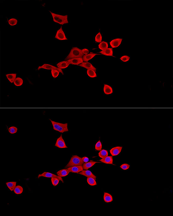
Immunofluorescence analysis of HepG2 cells using SLC27A2 Rabbit pAb (A1077) at dilution of 1:50 (40x lens). Secondary antibody: Cy3-conjugated Goat anti-Rabbit IgG (H+L) (AS007) at 1:500 dilution. Blue: DAPI for nuclear staining. |
|
|
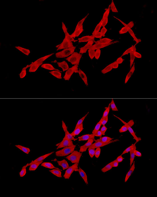
Immunofluorescence analysis of NIH/3T3 cells using SLC27A2 Rabbit pAb (A1077) at dilution of 1:50 (40x lens). Secondary antibody: Cy3-conjugated Goat anti-Rabbit IgG (H+L) (AS007) at 1:500 dilution. Blue: DAPI for nuclear staining. |
|
|
Immunofluorescence analysis of PC-12 cells using SLC27A2 Rabbit pAb (A1077) at dilution of 1:50 (40x lens). Secondary antibody: Cy3-conjugated Goat anti-Rabbit IgG (H+L) (AS007) at 1:500 dilution. Blue: DAPI for nuclear staining. |
Product Guarantee and Expert Support
