
GSDMD (Full length+N terminal) Rabbit pAb, Unconjugated, Polyclonal
Catalog Number:
ABB-A10164
- Images (7)

| Article Name: | GSDMD (Full length+N terminal) Rabbit pAb, Unconjugated, Polyclonal |
| Biozol Catalog Number: | ABB-A10164 |
| Supplier Catalog Number: | A10164 |
| Alternative Catalog Number: | ABB-A10164-100UL,ABB-A10164-20UL,ABB-A10164-1000UL,ABB-A10164-500UL |
| Manufacturer: | ABclonal |
| Host: | Rabbit |
| Category: | Antikörper |
| Application: | ELISA, IF, WB |
| Species Reactivity: | Mouse |
| Immunogen: | Recombinant protein (or fragment).This information is considered to be commercially sensitive. |
| Conjugation: | Unconjugated |
| Alternative Names: | DF5L, M2-4, Dfna5l, GsdmD-1, Gsdmdc1, 1810036L03Rik, GSDMD (Full Length+N terminal) |
| Enables phospholipid binding activity and wide pore channel activity. Involved in several processes, including defense response to bacterium, positive regulation of interleukin-1 beta production, and protein-containing complex assembly. Acts upstream of or within cellular response to extracellular stimulus. Located in cytosol, extracellular space, and plasma membrane. Part of NLRP3 inflammasome complex. Is expressed in several structures, including gut, liver, lung, metanephros, and spleen. Orthologous to human GSDMD (gasdermin D). |
| Application Dilute: | WB,1:100 - 1:500|IF/ICC,1:50 - 1:200|ELISA,Recommended starting concentration is 1 µg/mL. Please optimize the concentration based on your specific assay requirements. |
| Application Notes: | Cross-Reactivity: Human,Mouse,Rat. ResearchArea: Cell Biology & Developmental Biology. Shipping: Ice Bag |
|
|
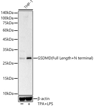
Western blot analysis of lysates from THP-1 cells, using GSDMD (Full Length+N terminal) Rabbit pAb (A10164) at 1:400 dilution. THP-1 cells were treated with PMA/TPA (80 nM) at 37°C for overnight and LPS (1 µg/ml) at 37°C for 6 hours Secondary antibody: HRP-conjugated Goat anti-Rabbit IgG (H+L) (AS014) at 1:10000 dilution. Lysates/proteins: 25µg per lane. Blocking buffer: 3% nonfat dry milk in TBST. Detection: ECL Basic Kit (RM00020). Exposure time: 1s. |
|
|
Western blot analysis of lysates from wild type (WT) and 293T transfected with GSDMD-N using GSDMD (Full Length+N terminal) (A10164) at 1:2000 dilution. Secondary antibody: HRP-conjugated Goat anti-Rabbit IgG (H+L) (AS014) at 1:10000 dilution. Lysates/proteins: 25µg per lane. Blocking buffer: 3% nonfat dry milk in TBST. Detection: ECL Basic Kit (RM00020). Exposure time: 1s. |
|
|
Western blot analysis of lysates from wild type (WT) and 293T transfected with GSDMD using GSDMD (Full Length+N terminal) (A10164) at 1:2000 dilution. Secondary antibody: HRP-conjugated Goat anti-Rabbit IgG (H+L) (AS014) at 1:10000 dilution. Lysates/proteins: 25µg per lane. Blocking buffer: 3% nonfat dry milk in TBST. Detection: ECL Basic Kit (RM00020). Exposure time: 0.5s. |
|
|
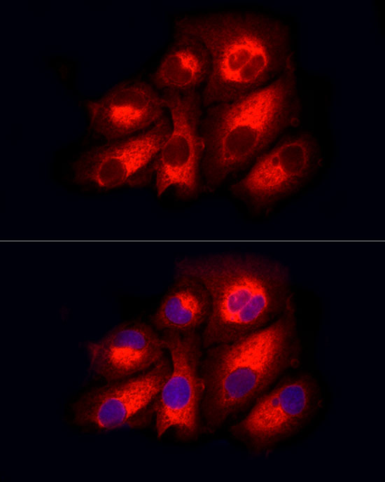
Immunofluorescence analysis of HeLa cells using GSDMD (Full Length+N terminal) Rabbit pAb (A10164) at dilution of 1:200 (40x lens). Secondary antibody: Cy3-conjugated Goat anti-Rabbit IgG (H+L) (AS007) at 1:500 dilution. Blue: DAPI for nuclear staining. |
|
|
Immunofluorescence analysis of NIH/3T3 cells using GSDMD (Full Length+N terminal) Rabbit pAb (A10164) at dilution of 1:200 (40x lens). Secondary antibody: Cy3-conjugated Goat anti-Rabbit IgG (H+L) (AS007) at 1:500 dilution. Blue: DAPI for nuclear staining. |
|
|
Immunofluorescence analysis of PC-12 cells using GSDMD (Full Length+N terminal) Rabbit pAb (A10164) at dilution of 1:200 (40x lens). Secondary antibody: Cy3-conjugated Goat anti-Rabbit IgG (H+L) (AS007) at 1:500 dilution. Blue: DAPI for nuclear staining. |
|
|
Immunofluorescence analysis of PC-3 cells using GSDMD (Full Length+N terminal) Rabbit pAb (A10164) at dilution of 1:200 (40x lens). Secondary antibody: Cy3-conjugated Goat anti-Rabbit IgG (H+L) (AS007) at 1:500 dilution. Blue: DAPI for nuclear staining. |
Product Guarantee and Expert Support
