
HSP60/HSPD1 Rabbit pAb, Unconjugated, Polyclonal
Catalog Number:
ABB-A0969
- Images (7)


| Article Name: | HSP60/HSPD1 Rabbit pAb, Unconjugated, Polyclonal |
| Biozol Catalog Number: | ABB-A0969 |
| Supplier Catalog Number: | A0969 |
| Alternative Catalog Number: | ABB-A0969-20UL,ABB-A0969-100UL,ABB-A0969-1000UL,ABB-A0969-500UL |
| Manufacturer: | ABclonal |
| Host: | Rabbit |
| Category: | Antikörper |
| Application: | ELISA, IF, IHC-P, IP, WB |
| Species Reactivity: | Human |
| Immunogen: | Recombinant protein (or fragment).This information is considered to be commercially sensitive. |
| Conjugation: | Unconjugated |
| Alternative Names: | HLD4, CPN60, GROEL, HSP60, HSP65, SPG13, HSP-60, HuCHA60, HSP60/HSPD1 |
| This gene encodes a member of the chaperonin family. The encoded mitochondrial protein may function as a signaling molecule in the innate immune system. This protein is essential for the folding and assembly of newly imported proteins in the mitochondria. This gene is adjacent to a related family member and the region between the 2 genes functions as a bidirectional promoter. Several pseudogenes have been associated with this gene. Two transcript variants encoding the same protein have been identified for this gene. Mutations associated with this gene cause autosomal recessive spastic paraplegia 13. |
| Clonality: | Polyclonal |
| Molecular Weight: | 61kDa |
| NCBI: | 3329 |
| UniProt: | P10809 |
| Purity: | Affinity purification |
| Sequence: | AKDVKFGADARALMLQGVDLLADAVAVTMGPKGRTVIIEQSWGSPKVTKDGVTVAKSIDLKDKYKNIGAKLVQDVANNTNEEAGDGTTTATVLARSIAKEGFEKISKGANPVEIRRGVMLAVDAVIAELKKQSKPVTTPEEIAQVATISANGDKEIGNIISDAMKKVGRKGVITVKDGKTLNDELEIIEGMKFDRGYISPYFINTSKGQKCEFQ |
| Target: | HSPD1 |
| Application Dilute: | WB,1:500 - 1:5000|IF/ICC,1:50 - 1:400|IHC-P,1:50 - 1:200|IP,0.5µg-4µg antibody for 200µg-400µg extracts of whole cells|ELISA,Recommended starting concentration is 1 µg/mL. Please optimize the concentration based on your specific assay requirements. |
| Application Notes: | Cross-Reactivity: Human,Mouse,Rat. ResearchArea: Epigenetics Nuclear Signaling,RNA Binding,Signal Transduction,Cell Biology Developmental Biology,Apoptosis,Mitochondrial Control of Apoptosis,Endocrine Metabolism,Mitochondrial metabolism. Shipping: Ice Bag |
|
|
Western blot analysis of various lysates using HSP60/HSPD1 Rabbit pAb (A0969) at 1:1000 dilution. Secondary antibody: HRP-conjugated Goat anti-Rabbit IgG (H+L) (AS014) at 1:10000 dilution. Lysates/proteins: 25µg per lane. Blocking buffer: 3% nonfat dry milk in TBST. Detection: ECL Basic Kit (RM00020). Exposure time: 10s. |
|
|
Immunohistochemistry analysis of paraffin-embedded Rat kidney using HSP60/HSPD1 Rabbit pAb (A0969) at dilution of 1:100 (40x lens). Microwave antigen retrieval performed with 0.01M Tris/EDTA Buffer (pH 9.0) prior to IHC staining. |
|
|
Immunohistochemistry analysis of paraffin-embedded Human colon using HSP60/HSPD1 Rabbit pAb (A0969) at dilution of 1:100 (40x lens). Microwave antigen retrieval performed with 0.01M Tris/EDTA Buffer (pH 9.0) prior to IHC staining. |
|
|
Immunohistochemistry analysis of paraffin-embedded Mouse testis using HSP60/HSPD1 Rabbit pAb (A0969) at dilution of 1:100 (40x lens). Microwave antigen retrieval performed with 0.01M Tris/EDTA Buffer (pH 9.0) prior to IHC staining. |
|
|
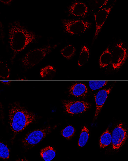
Confocal immunofluorescence analysis of HeLa cells using HSP60/HSPD1 Rabbit pAb (A0969) at dilution of 1:400. Blue: DAPI for nuclear staining. |
|
|
Confocal immunofluorescence analysis of U2OS cells using HSP60/HSPD1 Rabbit pAb (A0969) at dilution of 1:100. Blue: DAPI for nuclear staining. |
|
|
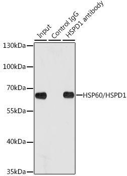
Immunoprecipitation analysis of 200 µg extracts of HeLa cells using 1 µg HSP60/HSPD1 antibody (A0969). Western blot was performed from the immunoprecipitate using HSP60/HSPD1 antibody (A0969) at a dilution of 1:1000. |
Product Guarantee and Expert Support
