
STAT6 Rabbit pAb, Unconjugated, Polyclonal
Catalog Number:
ABB-A0755
- Images (7)


| Article Name: | STAT6 Rabbit pAb, Unconjugated, Polyclonal |
| Biozol Catalog Number: | ABB-A0755 |
| Supplier Catalog Number: | A0755 |
| Alternative Catalog Number: | ABB-A0755-100UL,ABB-A0755-20UL,ABB-A0755-500UL,ABB-A0755-1000UL |
| Manufacturer: | ABclonal |
| Host: | Rabbit |
| Category: | Antikörper |
| Application: | ELISA, IF, WB |
| Species Reactivity: | Human |
| Immunogen: | Synthetic peptide. This information is considered to be commercially sensitive. |
| Conjugation: | Unconjugated |
| Alternative Names: | STAT6B, STAT6C, D12S1644, IL-4-STAT, STAT6 |
| The protein encoded by this gene is a member of the STAT family of transcription factors. In response to cytokines and growth factors, STAT family members are phosphorylated by the receptor associated kinases, and then form homo- or heterodimers that translocate to the cell nucleus where they act as transcription activators. This protein plays a central role in exerting IL4 mediated biological responses. It is found to induce the expression of BCL2L1/BCL-X(L), which is responsible for the anti-apoptotic activity of IL4. Knockout studies in mice suggested the roles of this gene in differentiation of T helper 2 (Th2) cells, expression of cell surface markers, and class switch of immunoglobulins. Alternative splicing results in multiple transcript variants. |
| Clonality: | Polyclonal |
| Molecular Weight: | 94kDa |
| NCBI: | 6778 |
| UniProt: | P42226 |
| Purity: | Affinity purification |
| Sequence: | QFNKEILLGRGFTFWQWFDGVLDLTKRCLRSYWSDRLIIGFISKQYVTSLLLNEPDGTFLLRFSDSEIGGITIAHVIRGQDGSPQIENIQPFSAKDLSIRSLGDRIRDLAQLKNLYPKKPKDEAFRSHYKPEQMGKDGRGYVPATIKMTVERDQPLPTPELQMPTMVPSYDLGMAPDSSMSMQLGPDMVPQVYPPHSHSIPPYQGLSPEESVNVLSAFQEPHLQMPPSLGQMSLPFDQPHPQGLLPCQPQEHAVS |
| Target: | STAT6 |
| Antibody Type: | Primary Antibody |
| Application Dilute: | WB,1:500 - 1:1000|IF/ICC,1:50 - 1:200|ELISA,Recommended starting concentration is 1 µg/mL. Please optimize the concentration based on your specific assay requirements. |
| Application Notes: | Cross-Reactivity: Human,Mouse,Rat. ResearchArea: Epigenetics Nuclear Signaling,Transcription Factors,Protein phosphorylation,Signal Transduction,Immunology Inflammation,Jak-Stat-IL-6 Receptor Signaling Pathway. Shipping: Ice Bag |
|
|
Immunofluorescence analysis of HeLa cells using STAT6 Rabbit pAb (A0755) at dilution of 1:200 (40x lens). Secondary antibody: Cy3-conjugated Goat anti-Rabbit IgG (H+L) (AS007) at 1:500 dilution. Blue: DAPI for nuclear staining. |
|
|
Immunofluorescence analysis of NIH/3T3 cells using STAT6 Rabbit pAb (A0755) at dilution of 1:200 (40x lens). Secondary antibody: Cy3-conjugated Goat anti-Rabbit IgG (H+L) (AS007) at 1:500 dilution. Blue: DAPI for nuclear staining. |
|
|
Western blot analysis of various lysates using STAT6 Rabbit pAb (A0755) at 1:1000 dilution. Secondary antibody: HRP-conjugated Goat anti-Rabbit IgG (H+L) (AS014) at 1:10000 dilution. Lysates/proteins: 25µg per lane. Blocking buffer: 3% nonfat dry milk in TBST. Detection: ECL Basic Kit (RM00020). Exposure time: 10s. |
|
|
Immunofluorescence analysis of PC-12 cells using STAT6 Rabbit pAb (A0755) at dilution of 1:200 (40x lens). Secondary antibody: Cy3-conjugated Goat anti-Rabbit IgG (H+L) (AS007) at 1:500 dilution. Blue: DAPI for nuclear staining. |
|
|
Immunofluorescence analysis of U2OS cells using STAT6 Rabbit pAb (A0755) at dilution of 1:200 (40x lens). Secondary antibody: Cy3-conjugated Goat anti-Rabbit IgG (H+L) (AS007) at 1:500 dilution. Blue: DAPI for nuclear staining. |
|
|
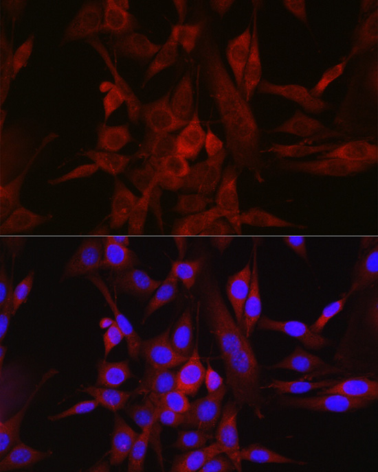
Immunofluorescence analysis of NIH/3T3 cells using STAT6 Rabbit pAb (A0755) at dilution of 1:100 (40x lens). Secondary antibody: Cy3-conjugated Goat anti-Rabbit IgG (H+L) (AS007) at 1:500 dilution. Blue: DAPI for nuclear staining. |
|
|
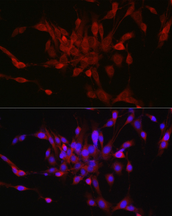
Immunofluorescence analysis of PC-12 cells using STAT6 Rabbit pAb (A0755) at dilution of 1:100 (40x lens). Secondary antibody: Cy3-conjugated Goat anti-Rabbit IgG (H+L) (AS007) at 1:500 dilution. Blue: DAPI for nuclear staining. |
Product Guarantee and Expert Support
