
[KO Validated] HK1 Rabbit mAb, Unconjugated, Monoclonal
Catalog Number:
ABB-A0533
- Images (7)


| Article Name: | [KO Validated] HK1 Rabbit mAb, Unconjugated, Monoclonal |
| Biozol Catalog Number: | ABB-A0533 |
| Supplier Catalog Number: | A0533 |
| Alternative Catalog Number: | ABB-A0533-100UL,ABB-A0533-20UL,ABB-A0533-500UL,ABB-A0533-1000UL |
| Manufacturer: | ABclonal |
| Host: | Rabbit |
| Category: | Antikörper |
| Application: | ELISA, IF, IHC-P, IP, WB |
| Species Reactivity: | Human |
| Immunogen: | Synthetic peptide. This information is considered to be commercially sensitive. |
| Conjugation: | Unconjugated |
| Alternative Names: | HK, HKD, HKI, HXK1, NMSR, RP79, HMSNR, HK1-ta, HK1-tb, HK1-tc, NEDVIBA, hexokinase, K1 |
| Hexokinases phosphorylate glucose to produce glucose-6-phosphate, the first step in most glucose metabolism pathways. This gene encodes a ubiquitous form of hexokinase which localizes to the outer membrane of mitochondria. Mutations in this gene have been associated with hemolytic anemia due to hexokinase deficiency. Alternative splicing of this gene results in several transcript variants which encode different isoforms, some of which are tissue-specific. |
| Application Dilute: | WB,1:1000 - 1:2000|IP,0.5µg-4µg antibody for 400µg-600µg extracts of whole cells|IF/ICC,1:200 - 1:800|IF-P,1:200 - 1:800|IHC-P,1:200 - 1:800|ELISA,Recommended starting concentration is 1 µg/mL. Please optimize the concentration based on your specific assa |
| Application Notes: | Cross-Reactivity: Human,Mouse,Rat. ResearchArea: Cancer,Signal Transduction,Endocrine Metabolism,Carbohydrate metabolism,Warburg Effect,Cardiovascular,Blood,Heart. Shipping: Ice Bag |
|
|
Western blot analysis of various lysates using [KO Validated] HK1 Rabbit mAb (A0533) at 1:1000 dilution. Secondary antibody: HRP-conjugated Goat anti-Rabbit IgG (H+L) (AS014) at 1:10000 dilution. Lysates/proteins: 25µg per lane. Blocking buffer: 3% nonfat dry milk in TBST. Detection: ECL Basic Kit (RM00020). Exposure time: 60s. |
|
|
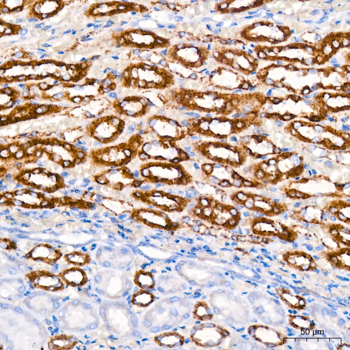
Immunohistochemistry analysis of paraffin-embedded Mouse kidney tissue using [KO Validated] HK1 Rabbit mAb (A0533) at dilution of 1:200 (40x lens). High pressure antigen retrieval performed with 0.01M Citrate buffer (pH 6.0) prior to IHC staining. |
|
|
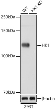
Western blot analysis of lysates from wild type (WT) and HK1 knockout (KO) 293T cells, using [KO Validated] HK1 Rabbit mAb (A0533) at 1:1000 dilution. Secondary antibody: HRP-conjugated Goat anti-Rabbit IgG (H+L) (AS014) at 1:10000 dilution. Lysates/proteins: 25µg per lane. Blocking buffer: 3% nonfat dry milk in TBST. Detection: ECL Basic Kit (RM00020). Exposure time: 60s. |
|
|
Immunohistochemistry analysis of paraffin-embedded Rat kidney tissue using [KO Validated] HK1 Rabbit mAb (A0533) at dilution of 1:200 (40x lens). High pressure antigen retrieval performed with 0.01M Citrate buffer (pH 6.0) prior to IHC staining. |
|
|
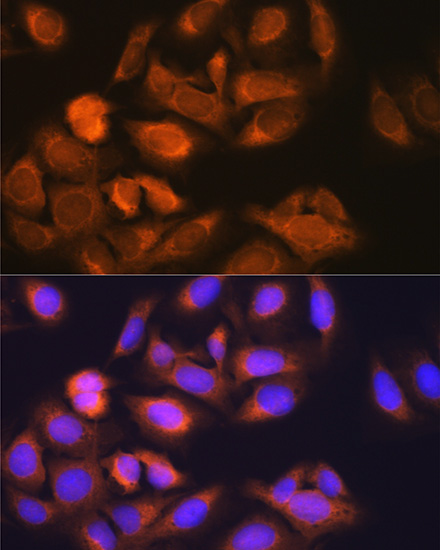
Confocal imaging of U-2 OS cells using [KO Validated] HK1 Rabbit mAb (A0533, dilution 1:200) followed by a further incubation with Cy3 Goat Anti-Rabbit IgG (H+L) (AS007, dilution 1:500) (Red). The cells were counterstained with alpha-Tubulin Mouse mAb (AC012, dilution 1:400) followed by incubation with ABflo 488-conjugated Goat Anti-Mouse IgG (H+L) Ab (AS076, dilution 1:500) (Green). DAPI was used for nuclear staining (Blue). Objective: 100x. |
|
|
Confocal imaging of paraffin-embedded Mouse kidney tissue using [KO Validated] HK1 Rabbit mAb (A0533, dilution 1:200) followed by a further incubation with Cy3 Goat Anti-Rabbit IgG (H+L) (AS007, dilution 1:500) (Red). DAPI was used for nuclear staining (Blue). Objective: 40x.Perform high pressure antigen retrieval with 0.01M citrate buffer (pH 6.0) prior to IF staining. |
|
|
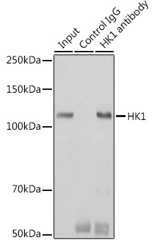
Immunoprecipitation analysis of 600 µg extracts of Mouse brain cells using 3 µg [KO Validated] HK1 Rabbit mAb (A0533). Western blot was performed from the immunoprecipitate using HK1 (A0533) at a dilution of 1:1000. |
Product Guarantee and Expert Support
