
IFIT3 Antibody, Unconjugated, Rabbit, Polyclonal
Artikelnummer:
CSB-PA011022HA01HU
- Bilder (5)
| Artikelname: | IFIT3 Antibody, Unconjugated, Rabbit, Polyclonal |
| Artikelnummer: | CSB-PA011022HA01HU |
| Hersteller Artikelnummer: | CSB-PA011022HA01HU |
| Alternativnummer: | CSB-PA011022HA01HU-100UG, CSB-PA011022HA01HU-50UG |
| Hersteller: | Cusabio |
| Wirt: | Rabbit |
| Kategorie: | Antikörper |
| Applikation: | ELISA, IF, IHC, IP, WB |
| Spezies Reaktivität: | Human |
| Konjugation: | Unconjugated |
| Alternative Synonym: | CIG 49 antibody, CIG49 antibody, GARG 49 antibody, IFI-60K antibody, IFI60 antibody, IFI60K antibody, IFIT-3 antibody, IFIT-4 antibody, Ifit3 antibody, IFIT3_HUMAN antibody, IFIT4 antibody, Interferon induced 60 kDa protein antibody, Interferon induced protein 60 antibody, Interferon induced protein with tetratricopeptide repeats 3 antibody, Interferon-induced 60 kDa protein antibody, Interferon-induced protein with tetratricopeptide repeats 3 antibody, Interferon-induced protein with tetratricopeptide repeats 4 antibody, IRG2 antibody, ISG-60 antibody, ISG60 antibody, P60 antibody, Retinoic acid induced gene G protein antibody, Retinoic acid-induced gene G protein antibody, RIG G antibody, RIG-G antibody, RIGG antibody, RP11-149I23.4 antibody |
| Klonalität: | Polyclonal |
| UniProt: | O14879 |
| Puffer: | Preservative: 0.03% Proclin 300<br />Constituents: 50% Glycerol, 0.01M PBS, PH 7.4 |
| Reinheit: | >95%, Protein G purified |
| Formulierung: | Liquid |
| Target-Kategorie: | IFIT3 |
| Application Verdünnung: | Recommended dilution: WB:1:1000-1:5000, IHC:1:20-1:200, IF:1:50-1:500, IP:1:200-1:2000 |
|
|
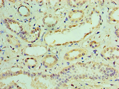
Immunohistochemistry of paraffin-embedded human kidney tissue using CSB-PA011022HA01HU at dilution of 1:100 |
|
|
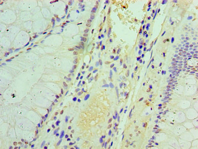
Immunohistochemistry of paraffin-embedded human colon cancer using CSB-PA011022HA01HU at dilution of 1:100 |
|
|
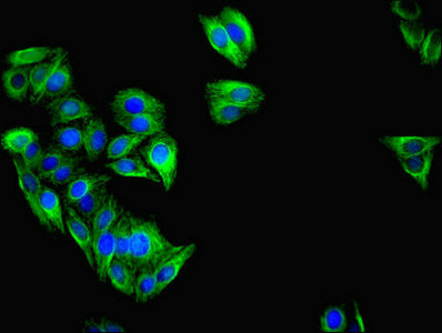
Immunofluorescent analysis of HepG2 cells using CSB-PA011022HA01HU at dilution of 1:100 and Alexa Fluor 488-congugated AffiniPure Goat Anti-Rabbit IgG(H+L) |
|
|
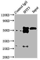
Immunoprecipitating IFIT3 in HepG2 whole cell lysate Lane 1: Rabbit control IgG (1µg) instead of CSB-PA011022HA01HU in HepG2 whole cell lysate. For western blotting, a HRP-conjugated Protein G antibody was used as the secondary antibody (1/2000) Lane 2: CSB-PA011022HA01HU (6µg) + HepG2 whole cell lysate (500µg) Lane 3: HepG2 whole cell lysate (10µg) |
|
|
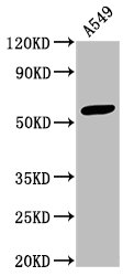
Western Blot Positive WB detected in: A549 whole cell lysate All lanes: IFIT3 antibody at 3.3µg/ml Secondary Goat polyclonal to rabbit IgG at 1/50000 dilution Predicted band size: 56 kDa Observed band size: 56 kDa |
Produktgarantie und fachkundiger Support
