
HIST1H3A (Ab-4) Antibody, Unconjugated, Rabbit, Polyclonal
Artikelnummer:
CSB-PA010418OA04NACHU
- Bilder (5)
| Artikelname: | HIST1H3A (Ab-4) Antibody, Unconjugated, Rabbit, Polyclonal |
| Artikelnummer: | CSB-PA010418OA04NACHU |
| Hersteller Artikelnummer: | CSB-PA010418OA04nacHU |
| Alternativnummer: | CSB-PA010418OA04NACHU-100UL, CSB-PA010418OA04NACHU-50UL |
| Hersteller: | Cusabio |
| Wirt: | Rabbit |
| Kategorie: | Antikörper |
| Applikation: | ChIP, ELISA, IF, IHC, WB |
| Spezies Reaktivität: | Human, Mouse |
| Konjugation: | Unconjugated |
| Alternative Synonym: | H3 histone family member E pseudogene antibody, H3 histone family, member A antibody, H3/A antibody, H31_HUMAN antibody, H3F3 antibody, H3FA antibody, Hist1h3a antibody, HIST1H3B antibody, HIST1H3C antibody, HIST1H3D antibody, HIST1H3E antibody, HIST1H3F antibody, HIST1H3G antibody, HIST1H3H antibody, HIST1H3I antibody, HIST1H3J antibody, HIST3H3 antibody, histone 1, H3a antibody, Histone cluster 1, H3a antibody, Histone H3 3 pseudogene antibody, Histone H3.1 antibody, Histone H3/a antibody, Histone H3/b antibody, Histone H3/c antibody, Histone H3/d antibody, Histone H3/f antibody, Histone H3/h antibody, Histone H3/i antibody, Histone H3/j antibody, Histone H3/k antibody, Histone H3/l antibody |
| Klonalität: | Polyclonal |
| UniProt: | P68431 |
| Puffer: | Preservative: 0.03% Proclin 300<br />Constituents: 50% Glycerol, 0.01M PBS, pH 7.4 |
| Reinheit: | Antigen Affinity Purified |
| Formulierung: | Liquid |
| Target-Kategorie: | HIST1H3A |
| Application Verdünnung: | Recommended dilution: WB:1:50-1:500, IHC:1:20-1:200, IF:1:1-1:10 |
|
|
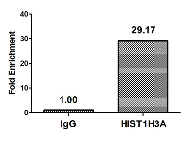
Chromatin Immunoprecipitation Hela (4*106) were treated with Micrococcal Nuclease, sonicated, and immunoprecipitated with 5µg anti-HIST1H3A (CSB-PA010418OA04nacHU) or a control normal rabbit IgG. The resulting ChIP DNA was quantified using real-time PCR with primers against the beta-Globin promoter. |
|
|
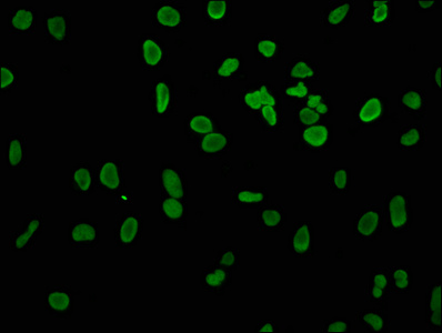
Immunofluorescence staining of Hela cells with CSB-PA010418OA04nacHU at 1:4, counter-stained with DAPI. The cells were fixed in 4% formaldehyde, permeabilized using 0.2% Triton X-100 and blocked in 10% normal Goat Serum. The cells were then incubated with the antibody overnight at 4°,C. The secondary antibody was Alexa Fluor 488-congugated AffiniPure Goat Anti-Rabbit IgG(H+L). |
|
|
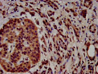
IHC image of CSB-PA010418OA04nacHU diluted at 1:100 and staining in paraffin-embedded human pancreatic cancer performed on a Leica BondTM system. After dewaxing and hydration, antigen retrieval was mediated by high pressure in a citrate buffer (pH 6.0). Section was blocked with 10% normal goat serum 30min at RT. Then primary antibody (1% BSA) was incubated at 4°,C overnight. The primary is detected by a biotinylated secondary antibody and visualized using an HRP conjugated SP system. |
|
|
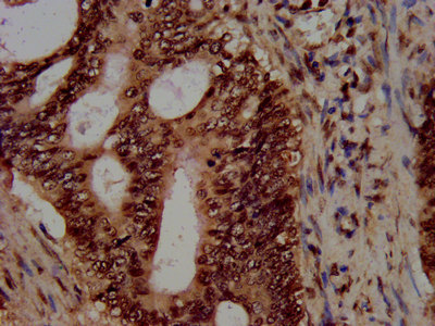
IHC image of CSB-PA010418OA04nacHU diluted at 1:100 and staining in paraffin-embedded human colon cancer performed on a Leica BondTM system. After dewaxing and hydration, antigen retrieval was mediated by high pressure in a citrate buffer (pH 6.0). Section was blocked with 10% normal goat serum 30min at RT. Then primary antibody (1% BSA) was incubated at 4°,C overnight. The primary is detected by a biotinylated secondary antibody and visualized using an HRP conjugated SP system. |
|
|
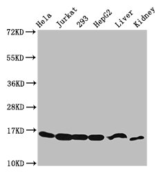
Western Blot Positive WB detected in: Hela whole cell lysate, Jurkat whole cell lysate, 293 whole cell lysate, HepG2 whole cell lysate, Mouse liver tissue, Mouse kidney tissue All lanes: HIST1H3A antibody at 2.48µg/ml Secondary Goat polyclonal to rabbit IgG at 1/50000 dilution Predicted band size: 16 kDa Observed band size: 16 kDa |
Produktgarantie und fachkundiger Support
