
HEPH Antibody, Unconjugated, Rabbit, Polyclonal
Artikelnummer:
CSB-PA010289ESR1HU
- Bilder (4)

| Artikelname: | HEPH Antibody, Unconjugated, Rabbit, Polyclonal |
| Artikelnummer: | CSB-PA010289ESR1HU |
| Hersteller Artikelnummer: | CSB-PA010289ESR1HU |
| Alternativnummer: | CSB-PA010289ESR1HU-100UL, CSB-PA010289ESR1HU-50UL |
| Hersteller: | Cusabio |
| Wirt: | Rabbit |
| Kategorie: | Antikörper |
| Applikation: | ELISA, IHC, WB |
| Spezies Reaktivität: | Human |
| Konjugation: | Unconjugated |
| Alternative Synonym: | CPL antibody, Heph antibody, HEPH_HUMAN antibody, Hephaestin antibody |
| Klonalität: | Polyclonal |
| UniProt: | Q9BQS7 |
| Puffer: | Preservative: 0.03% Proclin 300<br /> Constituents: 50% Glycerol, 0.01M PBS, PH 7.4 |
| Reinheit: | Antigen Affinity Purified |
| Formulierung: | Liquid |
| Target-Kategorie: | HEPH |
| Application Verdünnung: | Recommended dilution: WB:1:500-1:2000, IHC:1:20-1:100 |
|
|
|
|
|
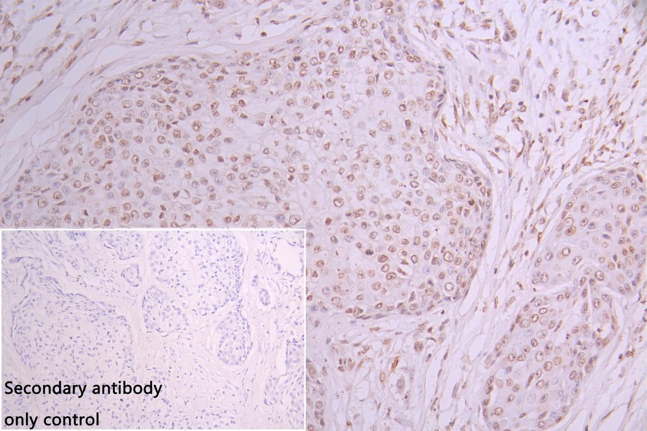
IHC image of CSB-PA010289ESR1HU diluted at 1:30 and staining in paraffin-embedded human pancreatic cancer performed on a Leica BondTM system. After dewaxing and hydration, antigen retrieval was mediated by high pressure in a citrate buffer (pH 6.0). Section was blocked with 10% normal goat serum 30min at RT. Then primary antibody (1% BSA) was incubated at 4C overnight. The primary is detected by a Goat anti-rabbit polymer IgG labeled by HRP and visualized using 0.05% DAB. Secondary antibody only control: uses 1% BSA instead of primary antibody |
|
|
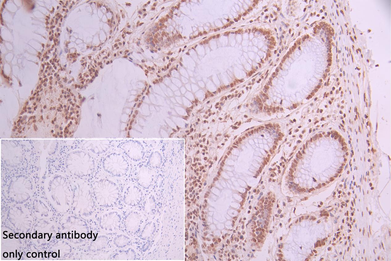
IHCimageofCSB-PA010289ESR1HUdilutedat1:30andstaininginparaffin-embeddedhumancolorectalcancerperformedonaLeicaBondTMsystem.Afterdewaxingandhydration,antigenretrievalwasmediatedbyhighpressureinacitratebuffer(pH6.0).Sectionwasblockedwith10%normalgoatserum30minatRT.Thenprimaryantibody(1%BSA)wasincubatedat4Covernight.TheprimaryisdetectedbyaGoatanti-rabbitpolymerIgGlabeledbyHRPandvisualizedusing0.05%DAB. Secondaryantibodyonlycontrol: uses 1%BSA instead of primary antibody |
|
|
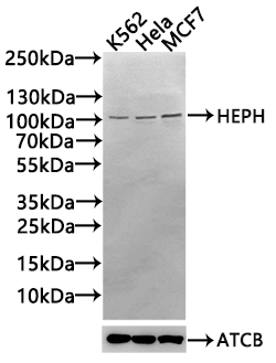
Western Blot Positive WB detected in: K562 whole cell lysate(30µg), Hela whole cell lysate(30µg), MCF7 whole cell lysate(30µg) All lanes: HEPH antibody at 1:1000 Secondary Goat polyclonal to rabbit IgG at 1/20000 dilution Predicted band size: 131,137,101 kDa Observed band size: 101 kDa Exposure time: 120s |
Produktgarantie und fachkundiger Support
