
GOT1 Antibody, Unconjugated, Rabbit, Polyclonal
Artikelnummer:
CSB-PA009679HA01HU
- Bilder (4)


| Artikelname: | GOT1 Antibody, Unconjugated, Rabbit, Polyclonal |
| Artikelnummer: | CSB-PA009679HA01HU |
| Hersteller Artikelnummer: | CSB-PA009679HA01HU |
| Alternativnummer: | CSB-PA009679HA01HU-100UG, CSB-PA009679HA01HU-50UG |
| Hersteller: | Cusabio |
| Wirt: | Rabbit |
| Kategorie: | Antikörper |
| Applikation: | ELISA, IF, IHC, WB |
| Spezies Reaktivität: | Human, Mouse |
| Konjugation: | Unconjugated |
| Alternative Synonym: | AATC_HUMAN antibody, Aspartate aminotransferase 1 antibody, Aspartate aminotransferase antibody, Aspartate aminotransferase cytoplasmic antibody, Aspartate aminotransferase cytosolic antibody, ASTQTL1 antibody, cAspAT antibody, cCAT antibody, cysteine aminotransferase, cytoplasmic antibody, cysteine transaminase, cytoplasmic antibody, cytoplasmic antibody, ec 2.6.1.1 antibody, GIG 18 antibody, GIG18 antibody, Glutamate oxaloacetate transaminase 1 antibody, Glutamate oxaloacetate transaminase soluble antibody, Glutamic oxaloacetic transaminase 1 soluble antibody, GOT 1 antibody, GOT1 antibody, Growth inhibiting protein 18 antibody, SGOT antibody, Transaminase A antibody |
| Klonalität: | Polyclonal |
| UniProt: | P17174 |
| Puffer: | Preservative: 0.03% Proclin 300<br />Constituents: 50% Glycerol, 0.01M PBS, PH 7.4 |
| Reinheit: | >95%, Protein G purified |
| Formulierung: | Liquid |
| Target-Kategorie: | GOT1 |
| Application Verdünnung: | Recommended dilution: WB:1:500-1:5000, IHC:1:200-1:500, IF:1:50-1:200 |
|
|
Western Blot Positive WB detected in: HepG2 whole cell lysate, Mouse liver tissue, Mouse skeletal muscle tissue All lanes: GOT1 antibody at 4.5µg/ml Secondary Goat polyclonal to rabbit IgG at 1/50000 dilution Predicted band size: 47, 45 kDa Observed band size: 47 kDa |
|
|
Immunofluorescence staining of HepG2 cells with CSB-PA009679HA01HU at 1:166, counter-stained with DAPI. The cells were fixed in 4% formaldehyde, permeabilized using 0.2% Triton X-100 and blocked in 10% normal Goat Serum. The cells were then incubated with the antibody overnight at 4°,C. The secondary antibody was Alexa Fluor 488-congugated AffiniPure Goat Anti-Rabbit IgG(H+L). |
|
|
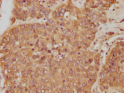
IHC image of CSB-PA009679HA01HU diluted at 1:250 and staining in paraffin-embedded human liver cancer performed on a Leica BondTM system. After dewaxing and hydration, antigen retrieval was mediated by high pressure in a citrate buffer (pH 6.0). Section was blocked with 10% normal goat serum 30min at RT. Then primary antibody (1% BSA) was incubated at 4°,C overnight. The primary is detected by a biotinylated secondary antibody and visualized using an HRP conjugated SP system. |
|
|
IHC image of CSB-PA009679HA01HU diluted at 1:250 and staining in paraffin-embedded human liver tissue performed on a Leica BondTM system. After dewaxing and hydration, antigen retrieval was mediated by high pressure in a citrate buffer (pH 6.0). Section was blocked with 10% normal goat serum 30min at RT. Then primary antibody (1% BSA) was incubated at 4°,C overnight. The primary is detected by a biotinylated secondary antibody and visualized using an HRP conjugated SP system. |
Produktgarantie und fachkundiger Support
