

CPT2 Recombinant Rabbit Monoclonal Antibody, Clone: [3B10], Unconjugated
Artikelnummer:
BYT-ORB612213
- Bilder (6)



| Artikelname: | CPT2 Recombinant Rabbit Monoclonal Antibody, Clone: [3B10], Unconjugated |
| Artikelnummer: | BYT-ORB612213 |
| Hersteller Artikelnummer: | orb612213 |
| Alternativnummer: | BYT-ORB612213-50,BYT-ORB612213-100 |
| Hersteller: | Biorbyt |
| Wirt: | Rabbit |
| Kategorie: | Antikörper |
| Applikation: | ICC, IF, IHC-Fr, IHC-P, WB |
| Spezies Reaktivität: | Human, Mouse, Rat |
| Immunogen: | A synthesized peptide derived from human CPT2 (600-658aa) |
| Konjugation: | Unconjugated |
| Alternative Synonym: | CPT1, CPTASE, IIAE4, CPTII, CPT2_HUMAN, CPT2, Carnitine palmitoyltransferase II (CPT II), 2.3.1.21, CPT2_MOUSE, Cpt-2, CPT2_RAT, |
| CPT2 Recombinant Rabbit Monoclonal Antibody |
| Klonalität: | Recombinant |
| Konzentration: | 1mg/ml |
| Klon-Bezeichnung: | [3B10] |
| Molekulargewicht: | 71 kDa |
| UniProt: | P23786 |
| Puffer: | 0.01M TBS (pH7.4) with 1% rAlbumin, 0.02% Proclin300 and 50% Glycerol. |
| Formulierung: | Liquid |
| Target-Kategorie: | CPT2 |
| Application Verdünnung: | WB=1:500-2000, IHC-P=1:50-200, IHC-F=1:50-200, ICC/IF=1:50-200, IF=1:50-200 |
|
|
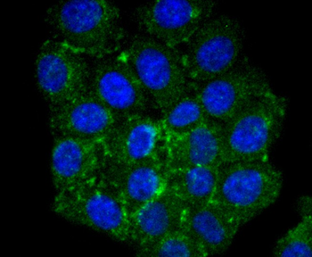
HepG2 cell, 4% Paraformaldehyde-fixed, Triton X-100 at room temperature for 20 min, Blocking buffer (normal goat serum) at 37C for 20 min, Antibody incubation with (CPT2) monoclonal Antibody, Unconjugated (orb612213) 1:50, 90 minutes at 37C, followed by a conjugated Goat Anti-Rabbit IgG antibody at 37C for 90 minutes, DAPI (blue) was used to stain the cell nuclei. |
|
|
HepG2 cell, 4% Paraformaldehyde-fixed, Triton X-100 at room temperature for 20 min, Blocking buffer (normal goat serum) at 37C for 20 min, Antibody incubation with (CPT2) monoclonal Antibody, Unconjugated (orb612213) 1:50, 90 minutes at 37C, followed by a conjugated Goat Anti-Rabbit IgG antibody at 37C for 90 minutes, DAPI (blue) was used to stain the cell nuclei. |
|
|
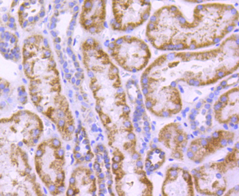
Paraformaldehyde-fixed, paraffin embedded (human kidney tissue), Antigen retrieval by boiling in sodium citrate buffer (pH6.0) for 15 min, Block endogenous peroxidase by 3% hydrogen peroxide for 20 minutes, Blocking buffer (normal goat serum) at 37C for 30 min, Antibody incubation with (CPT2) Monoclonal Antibody, Unconjugated (orb612213) at 1:50 overnight at 4C, followed by operating according to SP Kit (Rabbit) instructionsand DAB staining. |
|
|
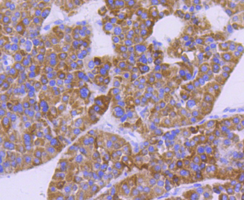
Paraformaldehyde-fixed, paraffin embedded (human liver carcinoma), Antigen retrieval by boiling in sodium citrate buffer (pH6.0) for 15 min, Block endogenous peroxidase by 3% hydrogen peroxide for 20 minutes, Blocking buffer (normal goat serum) at 37C for 30 min, Antibody incubation with (CPT2) Monoclonal Antibody, Unconjugated (orb612213) at 1:50 overnight at 4C, followed by operating according to SP Kit (Rabbit) instructionsand DAB staining. |
|
|
Paraformaldehyde-fixed, paraffin embedded (mouse kidney), Antigen retrieval by boiling in sodium citrate buffer (pH6.0) for 15 min, Block endogenous peroxidase by 3% hydrogen peroxide for 20 minutes, Blocking buffer (normal goat serum) at 37C for 30 min, Antibody incubation with (CPT2) Monoclonal Antibody, Unconjugated (orb612213) at 1:50 overnight at 4C, followed by operating according to SP Kit (Rabbit) instructionsand DAB staining. |
|
|
Sample: Lane 1: Hela cell lysate, Lane 2: 293 cell lysate, Lane 3: HepG2 cell lysate, Lane 4: NIH/3T3 cell lysate, Primary: Anti-CPT2 (orb612213) at 1:500 dilution, Secondary: Goat Anti-Rabbit IgG - HRP at 1:5000 dilution, Predicted band size: 71 kD, Observed band size: 71 kD. |
Produktgarantie und fachkundiger Support
