
Phospho-FLG (Tyr766) Rabbit Polyclonal Antibody, Unconjugated
Artikelnummer:
BYT-ORB6031
- Bilder (7)


| Artikelname: | Phospho-FLG (Tyr766) Rabbit Polyclonal Antibody, Unconjugated |
| Artikelnummer: | BYT-ORB6031 |
| Hersteller Artikelnummer: | orb6031 |
| Alternativnummer: | BYT-ORB6031-50,BYT-ORB6031-100,BYT-ORB6031-200 |
| Hersteller: | Biorbyt |
| Wirt: | Rabbit |
| Kategorie: | Antikörper |
| Applikation: | IF, IHC-Fr, IHC-P, WB |
| Spezies Reaktivität: | Human, Mouse, Rat |
| Immunogen: | KLH conjugated Synthesised phosphopeptide derived from human FGFR1 around the phosphorylation site of Tyr766 QE(p-Y)LD |
| Konjugation: | Unconjugated |
| Alternative Synonym: | FGFR1 (p-Y766), p-FGFR1, phospho-FGFR1, BFGFR, CD331, CEK, ECCL, FGFBR, FGFR-1, FLG, FLT-2, FLT2, HBGFR, HH2, HRTFDS, KAL2, N-SAM, OGD, bFGF-R-1, Eask, FGFR-I, Fr1, Hspy, MFR, c-fgr, bFGF-R, FGFR1_HUMAN, FGFR1, Basic fibroblast growth factor receptor 1 (BFGFR | bFGF-R-1), Fms-like tyrosine kinase 2 (FLT-2), Proto-oncogene c-Fgr, 2.7.10.1, FGFR1_MOUSE, Basic fibroblast growth factor receptor 1, FGFR1_RAT, |
| Phospho-FLG (Tyr766) Rabbit Polyclonal Antibody |
| Klonalität: | Polyclonal |
| Konzentration: | 1mg/ml |
| Molekulargewicht: | 88 kDa |
| UniProt: | P11362 |
| Puffer: | 0.01M TBS (pH7.4) with 1% rAlbumin, 0.02% Proclin300 and 50% Glycerol. |
| Formulierung: | Liquid |
| Target-Kategorie: | FGFR1 |
| Application Verdünnung: | WB=1:500-2000, IHC-P=1:100-500, IHC-F=1:100-500, IF=1:100-500 |
| Anwendungsbeschreibung: | Modification: Phosphorylated |
|
|
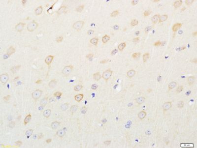
Immunohistochemical staining of rat brain tissue using FGF Receptor 1 (phospho-Tyr766) antibody. |
|
|
Western blot analysis of mouse Skin lysates (Lane 1) using FGF Receptor 1 (phospho-Tyr766) antibody. |
|
|
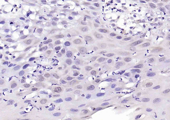
Paraformaldehyde-fixed, paraffin embedded (human laryngeal carcinoma), Antigen retrieval by boiling in sodium citrate buffer (pH6.0) for 15 min, Block endogenous peroxidase by 3% hydrogen peroxide for 20 minutes, Blocking buffer (normal goat serum) at 37C for 30 min, Antibody incubation with (Phospho-FLG (Tyr766)) Polyclonal Antibody, Unconjugated (orb6031) at 1:200 overnight at 4C, followed by operating according to SP Kit (Rabbit) instructionsand DAB staining. |
|
|
Paraformaldehyde-fixed, paraffin embedded (mouse brain), Antigen retrieval by boiling in sodium citrate buffer (pH6.0) for 15 min, Block endogenous peroxidase by 3% hydrogen peroxide for 20 minutes, Blocking buffer (normal goat serum) at 37C for 30 min, Antibody incubation with (Phospho-FLG (Tyr766)) Polyclonal Antibody, Unconjugated (orb6031) at 1:200 overnight at 4C, followed by operating according to SP Kit (Rabbit) instructionsand DAB staining. |
|
|
Sample: Cerebrum (Mouse) Lysate at 40 ug, Primary: Anti-Phospho-FLG (Tyr766) (orb6031) at 1/300 dilution, Secondary: IRDye800CW Goat Anti-Rabbit IgG at 1/20000 dilution, Predicted band size: 88 kD, Observed band size: 72 kD. |
|
|
Tissue/Cell: rat brain tissue, 4% Paraformaldehyde-fixed and paraffin-embedded, Antigen retrieval: citrate buffer (0.01M, pH6.0), Boiling bathing for 15 min, Block endogenous peroxidase by 3% Hydrogen peroxide for 30 min, Blocking buffer (normal goat serum) at 37C for 20 min, Incubation: Anti-Phospho-FLG (Tyr766) Polyclonal Antibody, Unconjugated (orb6031) 1:400, overnight at 4C, followed by conjugation to the secondary antibody and DAB staining. |
|
|
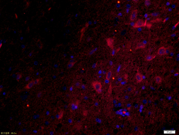
Tissue/Cell: rat brain tissue, 4% Paraformaldehyde-fixed and paraffin-embedded, Antigen retrieval: citrate buffer (0.01M, pH6.0), Boiling bathing for 15 min, Blocking buffer (normal goat serum) at 37C for 20 min, Incubation: Anti-p-FGFR1 Polyclonal Antibody, Unconjugated (orb6031) 1:200, overnight at 4C, The secondary antibody was Goat Anti-Rabbit IgG, Cy3 conjugated (orb868589) used at 1:200 dilution for 40 minutes at 37C. DAPI (5 ug/ml, blue) was used to stain the cell nuclei. |
Produktgarantie und fachkundiger Support
