A synthetic peptide corresponding to a sequence in the middle region of human GSTM1, which shares 70.6% and 73.5% amino acid (aa) sequence identity with mouse and rat GSTM1, respectively.
Konjugation:
Unconjugated
Alternative Synonym:
glutathione S transferase mu 1, GST class mu 1, GST HB subunit 4, GST1, GSTM1, GSTM1 1, GSTM1a 1a, GSTM1b 1b, GTH4, GTM1, H B, MU, MU 1
Anti-GSTM1 Antibody (monoclonal, 11F2). Tested in Flow Cytometry, IHC, ICC, WB applications. This antibody reacts with Human, Mouse, Rat.
Klonalität:
Monoclonal
Konzentration:
Adding 0.2 ml of distilled water will yield a concentration of 500 µg/ml.
Flow Cytometry analysis of HELA cells using anti-GSTM1 antibody. Overlay histogram showing HELA cells (Blue line). To facilitate intracellular staining, cells were fixed with 4% paraformaldehyde and permeabilized with permeabilization buffer. The cells were blocked with 10% normal goat serum. And then incubated with mouse anti-GSTM1 Antibody (1 µg/1x10 6 cells) for 30 min at 20C. DyLight488 conjugated goat anti-mouse IgG (5-10 µg/1x10 6 cells) was used as secondary antibody for 30 minutes at 20C. Isotype control antibody (Green line) was mouse IgG (1 µg/1x10 6) used under the same conditions. Unlabelled sample (Red line) was also used as a control.
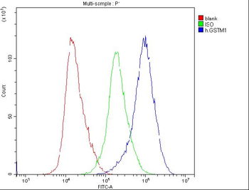
Flow Cytometry analysis of U20S cells using anti-GSTM1 antibody (Blu
Flow Cytometry analysis of U20S cells using anti-GSTM1 antibody. Overlay histogram showing U20S cells (Blue line). To facilitate intracellular staining, cells were fixed with 4% paraformaldehyde and permeabilized with permeabilization buffer. The cells were blocked with 10% normal goat serum. And then incubated with mouse anti-GSTM1 Antibody (1 µg/1x10 6 cells) for 30 min at 20C. DyLight488 conjugated goat anti-mouse IgG (5-10 µg/1x10 6 cells) was used as secondary antibody for 30 minutes at 20C. Isotype control antibody (Green line) was mouse IgG (1 µg/1x10 6) used under the same conditions. Unlabelled sample (Red line) was also used as a control.
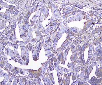
IHC analysis of GSTM1 using anti-GSTM1 antibody. GSTM1 was detected in paraffin-embedded section of human colon cancer tissue. Heat mediated antigen retrieval was performed in citrate buffer (pH6, epitope retrieval solution) for 20 mins. The tissue section was blocked with 10% goat serum. The tissue section was then incubated with 2 µg/ml mouse anti-GSTM1 Antibody overnight at 4C. Biotinylated goat anti-mouse IgG was used as secondary antibody and incubated for 30 minutes at 37C. The tissue section was developed using Strepavidin-Biotin-Complex (SABC) with DAB as the chromogen.
IHC analysis of GSTM1 using anti-GSTM1 antibody. GSTM1 was detected in paraffin-embedded section of rat liver tissue. Heat mediated antigen retrieval was performed in citrate buffer (pH6, epitope retrieval solution) for 20 mins. The tissue section was blocked with 10% goat serum. The tissue section was then incubated with 2 µg/ml mouse anti-GSTM1 Antibody overnight at 4C. Biotinylated goat anti-mouse IgG was used as secondary antibody and incubated for 30 minutes at 37C. The tissue section was developed using Strepavidin-Biotin-Complex (SABC) with DAB as the chromogen.
Western blot analysis of GSTM1 using anti-GSTM1 antibody. Electrophoresis was performed on a 5-20% SDS-PAGE gel at 70V (Stacking gel) / 90V (Resolving gel) for 2-3 hours. The sample well of each lane was loaded with 50 ug of sample under reducing conditions. Lane 1: human Hela, whole cell lysate, Lane 2: human T-47D whole cell lysate, Lane 3: rat brain tissue lysate, Lane 4: rat lung tissue lysate, Lane 5: rat stomach tissue lysate, Lane 6: mouse lung tissue lysate, Lane 7: mouse stomach tissue lysate, Lane 8: mouse kidney tissue lysate. After Electrophoresis, proteins were transferred to a Nitrocellulose membrane at 150mA for 50-90 minutes. Blocked the membrane with 5% Non-fat Milk/ TBS for 1.5 hour at RT. The membrane was incubated with mouse anti-GSTM1 antigen affinity purified monoclonal antibody at 0.5 µg/mL overnight at 4C, then washed with TBS-0.1% Tween 3 times with 5 minutes each and probed with a goat anti-mouse IgG-HRP secondary antibody at a dilution of 1:10000 for 1.5 hour at RT. The signal is developed using an Enhanced Chemiluminescent detection (ECL) kit with Tanon 5200 system. A specific band was detected for GSTM1 at approximately 26KD. The expected band size for GSTM1 is at 26KD.
* Mehrwertsteuer und Versandkosten nicht enthalten. Irrtümer und Preisänderungen vorbehalten