Cytochrome C CYCS Mouse Monoclonal Antibody, Clone: [15F10], Unconjugated
Artikelnummer:
BYT-ORB443140
Hersteller Artikelnummer:
orb443140
Alternativnummer:
BYT-ORB443140-100
Hersteller:
Biorbyt
Wirt:
Mouse
Kategorie:
Antikörper
Applikation:
FC, ICC, IF, IHC, WB
Spezies Reaktivität:
Human, Mouse, Rat
Immunogen:
E.coli-derived human Cytochrome C recombinant protein (Position: G2-E105). Human Cytochrome C shares 91% amino acid (aa) sequence identity with both mouse and rat Cytochrome C.
Anti-Cytochrome C CYCS Antibody (monoclonal, 15F10). Tested in Flow Cytometry, IF, IHC, ICC, WB applications. This antibody reacts with Human, Mouse, Rat.
Klonalität:
Monoclonal
Konzentration:
Adding 0.2 ml of distilled water will yield a concentration of 500 µg/ml.
Western blot, 0.1-0.5µg/ml Immunohistochemistry (Paraffin-embedded Section), 0.5-1µg/ml Immunocytochemistry/Immunofluorescence, 2µg/ml, Human Flow Cytometry (Fixed), 1-3µg/1x106 cells
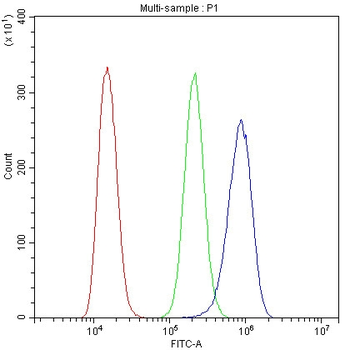
Flow Cytometry analysis of A431 cells using anti-Cytochrome C antibody. Overlay histogram showing A431 cells (Blue line). To facilitate intracellular staining, cells were fixed with 4% paraformaldehyde and permeabilized with permeabilization buffer. The cells were blocked with 10% normal goat serum. And then incubated with mouse anti-Cytochrome C Antibody (1 µg/1x10 6 cells) for 30 min at 20C. DyLight488 conjugated goat anti-mouse IgG (5-10 µg/1x10 6 cells) was used as secondary antibody for 30 minutes at 20C. Isotype control antibody (Green line) was mouse IgG (1 µg/1x10 6) used under the same conditions. Unlabelled sample (Red line) was also used as a control.
Flow Cytometry analysis of A431 cells using anti-Cytochrome C
IF analysis of Cytochrome C using anti-Cytochrome C antibody. Cytochrome C was detected in immunocytochemical section of MCF7 cells. Enzyme antigen retrieval was performed using IHC enzyme antigen retrieval reagent for 15 mins. The cells were blocked with 10% goat serum. And then incubated with 2 µg/mL mouse anti-Cytochrome C Antibody overnight at 4C. DyLight488 Conjugated Goat Anti-Mouse IgG was used as secondary antibody at 1:100 dilution and incubated for 30 minutes at 37C. The section was counterstained with DAPI. Visualize using a fluorescence microscope and filter sets appropriate for the label used.
IHC analysis of Cytochrome C using anti-Cytochrome C antibody. Cytochrome C was detected in paraffin-embedded section of human intestinal cancer tissue. Heat mediated antigen retrieval was performed in citrate buffer (pH6, epitope retrieval solution) for 20 mins. The tissue section was blocked with 10% goat serum. The tissue section was then incubated with 2 µg/ml mouse anti-Cytochrome C Antibody overnight at 4C. Biotinylated goat anti-mouse IgG was used as secondary antibody and incubated for 30 minutes at 37C. The tissue section was developed using Strepavidin-Biotin-Complex (SABC) with DAB as the chromogen.
IHC analysis of Cytochrome C using anti-Cytochrome C antibody. Cytochrome C was detected in paraffin-embedded section of human lung cancer tissue. Heat mediated antigen retrieval was performed in citrate buffer (pH6, epitope retrieval solution) for 20 mins. The tissue section was blocked with 10% goat serum. The tissue section was then incubated with 2 µg/ml mouse anti-Cytochrome C Antibody overnight at 4C. Biotinylated goat anti-mouse IgG was used as secondary antibody and incubated for 30 minutes at 37C. The tissue section was developed using Strepavidin-Biotin-Complex (SABC) with DAB as the chromogen.
IHC analysis of Cytochrome C using anti-Cytochrome C antibody. Cytochrome C was detected in paraffin-embedded section of human mammary cancer tissue. Heat mediated antigen retrieval was performed in citrate buffer (pH6, epitope retrieval solution) for 20 mins. The tissue section was blocked with 10% goat serum. The tissue section was then incubated with 2 µg/ml mouse anti-Cytochrome C Antibody overnight at 4C. Biotinylated goat anti-mouse IgG was used as secondary antibody and incubated for 30 minutes at 37C. The tissue section was developed using Strepavidin-Biotin-Complex (SABC) with DAB as the chromogen.
Western blot analysis of Cytochrome C using anti-Cytochrome C antibody. Electrophoresis was performed on a 12% SDS-PAGE gel at 70V (Stacking gel) / 90V (Resolving gel) for 2-3 hours. The sample well of each lane was loaded with 50 ug of sample under reducing conditions. Lane 1: human Hela whole cell lysate, Lane 2: human HepG2 whole cell lysate Lane 3: human K562 whole cell lysate, Lane 4: human Caco-2 whole cell lysate. After Electrophoresis, proteins were transferred to a Nitrocellulose membrane at 150mA for 50-90 minutes. Blocked the membrane with 5% Non-fat Milk/ TBS for 1.5 hour at RT. The membrane was incubated with mouse anti-Cytochrome C antigen affinity purified monoclonal antibody at 0.5 µg/mL overnight at 4C, then washed with TBS-0.1% Tween 3 times with 5 minutes each and probed with a goat anti-mouse IgG-HRP secondary antibody at a
* Mehrwertsteuer und Versandkosten nicht enthalten. Irrtümer und Preisänderungen vorbehalten