E.coli-derived human PKC alpha recombinant protein (Position: M153-L342). Human PKC alpha shares 97% and 99% amino acid (aa) sequence identity with mouse and rat PKC alpha, respectively.
Konjugation:
Unconjugated
Alternative Synonym:
Protein kinase C alpha type, PKC-A, PKC-alpha, 2.7.11.13, PRKCA, PKCA, PRKACA
PKC alpha/PRKCA Rabbit Polyclonal Antibody
Klonalität:
Polyclonal
Konzentration:
Adding 0.2 ml of distilled water will yield a concentration of 500 µg/ml.
Each vial contains antibody formulated with stabilizing components, 0.9 mg NaCl, 0.2 mg Na2HPO4, and 0.05 mg NaN3. *This antibody is supplied in a stabilized formulation. Compatibility with conjugation reactions depends on the chemistry of the conjugation
Formulierung:
Lyophilized
Target-Kategorie:
Protein kinase C alpha type
Application Verdünnung:
Western blot, 0.1-0.5µg/ml, Human, Mouse, Rat Immunohistochemistry (Paraffin-embedded Section), 0.5-1µg/ml, Human, Mouse, Rat
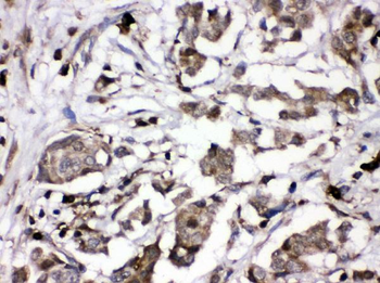
IHC analysis of PKC alpha using anti-PKC alpha antibody. PKC alpha was detected in paraffin-embedded section of human mammary cancer tissues. Heat mediated antigen retrieval was performed in citrate buffer (pH6, epitope retrieval solution) for 20 mins. The tissue section was blocked with 10% goat serum. The tissue section was then incubated with 1 µg/ml rabbit anti-PKC alpha Antibody overnight at 4C. Biotinylated goat anti-rabbit IgG was used as secondary antibody and incubated for 30 minutes at 37C. The tissue section was developed using Strepavidin-Biotin-Complex (SABC) with DAB as the chromogen.
IHC analysis of PKC alpha using anti-PKC alpha antibody.PKC alpha was detected
IHC analysis of PKC alpha using anti-PKC alpha antibody. PKC alpha was detected in paraffin-embedded section of mouse intestine tissues. Heat mediated antigen retrieval was performed in citrate buffer (pH6, epitope retrieval solution) for 20 mins. The tissue section was blocked with 10% goat serum. The tissue section was then incubated with 1 µg/ml rabbit anti-PKC alpha Antibody overnight at 4C. Biotinylated goat anti-rabbit IgG was used as secondary antibody and incubated for 30 minutes at 37C. The tissue section was developed using Strepavidin-Biotin-Complex (SABC) with DAB as the chromogen.
IHC analysis of PKC alpha using anti-PKC alpha antibody. PKC alpha was detected in paraffin-embedded section of rat intestine tissues. Heat mediated antigen retrieval was performed in citrate buffer (pH6, epitope retrieval solution) for 20 mins. The tissue section was blocked with 10% goat serum. The tissue section was then incubated with 1 µg/ml rabbit anti-PKC alpha Antibody overnight at 4C. Biotinylated goat anti-rabbit IgG was used as secondary antibody and incubated for 30 minutes at 37C. The tissue section was developed using Strepavidin-Biotin-Complex (SABC) with DAB as the chromogen.
Western blot analysis of PKC alpha using anti-PKC alpha antibody. Electrophoresis was performed on a 5-20% SDS-PAGE gel at 70V (Stacking gel) / 90V (Resolving gel) for 2-3 hours. The sample well of each lane was loaded with 30 ug of sample under reducing conditions. Lane 1: human U87 whole cell lysates, Lane 2: human HEL whole cell lysates, Lane 3: human A549 whole cell lysates, Lane 4: human U20S whole cell lysates, Lane 5: rat brain tissue lysates, Lane 6: mouse brain tissue lysates. After electrophoresis, proteins were transferred to a nitrocellulose membrane at 150 mA for 50-90 minutes. Blocked the membrane with 5% non-fat milk/TBS for 1.5 hour at RT. The membrane was incubated with rabbit anti-PKC alpha antigen affinity purified polyclonal antibody at 0.5 µg/mL overnight at 4C, then washed with TBS-0.1% Tween 3 times with 5 minutes each and probed with a goat anti-rabbit IgG-HRP secondary antibody at a dilution of 1:5000 for 1.5 hour at RT. The signal is developed using an Enhanced Chemiluminescent detection (ECL) kit with Tanon 5200 system. A specific band was detected for PKC alpha at approximately 77 kDa. The expected band size for PKC alpha is at 80 kDa.
Western blot analysis of PKC alpha/PRKCA using anti-PKC alpha/PRKCA Antibody. Electrophoresis was performed on a 5-20% SDS-PAGE gel at 70V (Stacking gel) / 90V (Resolving gel) for 2-3 hours. The sample well of each lane was loaded with 30 ug of sample under reducing conditions. After electrophoresis, proteins were transferred to a nitrocellulose membrane at 150 mA for 50-90 minutes. Blocked the membrane with 2.5% BSA on 1X-TBST 1.5 hour at RT. The membrane was incubated with rabbit anti-PKC alpha/PRKCA Antibody at 1:1000 (in 0.5%l BSA containing 1X-TBST) and overnight at 4C, then washed with TBS-0.1% Tween 3 times with 5 minutes each and probed with a donkey anti-rabbit Ig-Cy5 secondary antibody at a dilution of 1:1000 (in 0.5%l BSA containing 1X-TBST) at 25C for 2 h. The signal is developed using an CyDye. A specific band was detected for PKC alpha/PRKCA at approximately 76 kDa. The expected band size for PKC alpha/PRKCA is at 76 kDa.
* Mehrwertsteuer und Versandkosten nicht enthalten. Irrtümer und Preisänderungen vorbehalten