Purified polyclonal antibody supplied in PBS with 0.09% (W/V) sodium azide. This antibody is purified through a protein A column, followed by peptide affinity purification.
Application Verdünnung:
IHC-P - 1:100-500, FC - 1:25, WB - 1:2000
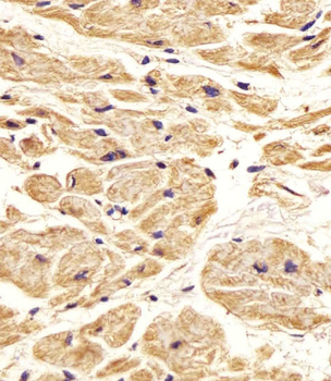
Staining Nestin in Human heart tissue sections by Immunohistochemistry (IHC-P - paraformaldehyde-fixed, paraffin-embedded sections). Tissue was fixed with formaldehyde and blocked with 3% BSA for 0.5 hour at room temperature, antigen retrieval was by heat mediation with a citrate buffer (pH6). Samples were incubated with primary antibody (1/25) for 1 hours at 37C. A undiluted biotinylated goat polyvalent antibody was used as the secondary antibody.
Staining Nestin in human brain tissue sections by Immunohistochemistry (IHC-P - paraformaldehyde-fixed, paraffin-embedded sections). Tissue was fixed with formaldehyde and blocked with 3% BSA for 0.5 hour at room temperature, antigen retrieval was by heat mediation with a citrate buffer (pH6). Samples were incubated with primary antibody (1/25) for 1 hours at 37C. A undiluted biotinylated goat polyvalent antibody was used as the secondary antibody.
Fluorescent confocal image of SH-SY5Y cells stained with Nestin (S1409) antibody. SH-SY5Y cells were fixed with 4% PFA (20 min), permeabilized with Triton X-100 (0.2%, 30 min). Cells were then incubated with Nestin (S1409) primary antibody (1:200, 2 h at room temperature). For secondary antibody, Alexa Fluor 488 conjugated donkey anti-rabbit antibody (green) was used (1:1000, 1 h). Nuclei were counterstained with Hoechst 33342 (blue) (10 µg/ml, 5 min).
Staining Nestin in human cerebellum tissue sections by Immunohistochemistry (IHC-P - paraformaldehyde-fixed, paraffin-embedded sections). Tissue was fixed with formaldehyde and blocked with 3% BSA for 0.5 hour at room temperature, antigen retrieval was by heat mediation with a citrate buffer (pH6). Samples were incubated with primary antibody (1/25) for 1 hours at 37C. A undiluted biotinylated goat polyvalent antibody was used as the secondary antibody.
Immunofluorescent analysis of 4% paraformaldehyde-fixed, 0.1% Triton X-100 permeabilized U-2 OS (human bone osteosarcoma cell line) cells labeling Nestin at 1/25 dilution, followed by Dylight 488-conjugated goat anti-rabbit IgG secondary antibody at 1/200 dilution (green). Immunofluorescence image showing cytoskeleton staining on U-2 OS cell line. Cytoplasmic actin is detected with Dylight 554 Phalloidin at 1/100 dilution (red).The nuclear counter stain is DAPI (blue).
Anti-Nestin Antibody (S1409) at 1:2000 dilution + Hela whole cell lysate. Lysates/proteins at 20 µg per lane. Secondary Goat Anti-Rabbit IgG, (H+L), Peroxidase conjugated at 1/10000 dilution. Predicted band size: 177 kDa. Blocking/Dilution buffer: 5% NFDM/TBST.
All lanes: Anti-Nestin Antibody (S1409) at 1:2000 dilution. Lane 1: U-251 MG whole cell lysate. Lane 2: SH-SY5Y whole cell lysate. Lysates/proteins at 20 µg per lane. Secondary Goat Anti-Rabbit IgG, (H+L), Peroxidase conjugated at 1/10000 dilution. Predicted band size: 177 kDa. Blocking/Dilution buffer: 5% NFDM/TBST.
* Mehrwertsteuer und Versandkosten nicht enthalten. Irrtümer und Preisänderungen vorbehalten