anti HRH2 antibody, anti histamine receptor H2 antibody, anti HGNC:5183 antibody, anti H2R antibody, anti gastric receptor 1 antibody, anti OTTHUMP00000161242 antibody
Supplied at 0.5 mg/ml in Tris saline, 0.02% sodium azide, pH 7.3 with 0.5% bovine serum albumin. Aliquot and store at -20C. Minimize freezing and thawing.
Sequenz:
QEEKPLKLQVWSGTE
Target-Kategorie:
Histamine Receptor H2
Application Verdünnung:
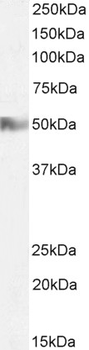
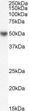
Peptide ELISA: antibody detection limit dilution 1:32000. Western blot: Approx 50kDa band observed in Human Tonsil lysates (calculated MW of 46.9kDa according to Human NP_001354640.1). Recommended concentration: 1-3µg/ml. Primary incubation 1 hour at room
2 µg/ml staining of Human Tonsil lysate (35 µg protein in RIPA buffer). Detected by chemiluminescence.
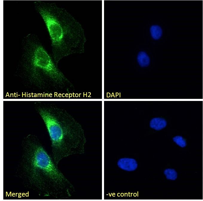
Immunofluorescence analysis of paraformaldehyde fixed HeLa cells, permeabilized with 0.15% Triton. Primary incubation 1hr (10 ug/ml) followed by Alexa Fluor 488 secondary antibody (2 ug/ml), showing cytoplasmic staining. The nuclear stain is DAPI (blue). Negative control: Unimmunized goat IgG (10 ug/ml) followed by Alexa Fluor 488 secondary antibody (2 ug/ml).
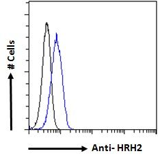
Flow cytometric analysis of paraformaldehyde fixed HeLa cells (blue line), permeabilized with 0.5% Triton. Primary incubation 1hr (10 ug/ml) followed by Alexa Fluor 488 secondary antibody (1 ug/ml). IgG control: Unimmunized goat IgG (black line) followed by Alexa Fluor 488 secondary antibody.
Western blot analysis of Human Tonsil lysate using HRH2 antibody
Immunofluorescence analysis of HeLa cells of HRH2 antibody
Flow cytometric analysis of HeLa cells using HRH2 antibody.
* Mehrwertsteuer und Versandkosten nicht enthalten. Irrtümer und Preisänderungen vorbehalten