Anti-RSPO1 antibody (orb1239947) was raised against a peptide corresponding to 16 amino acids near the amino terminus of human RSPO1. The immunogen is located within the first 50 amino acids of RSPO1.
RSPO1 Antibody is supplied in PBS containing 0.02% sodium azide.
Formulierung:
Liquid
Target-Kategorie:
RSPO1
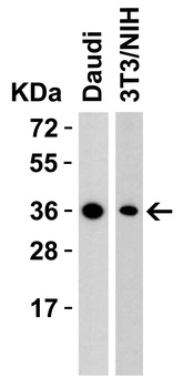
Western Blot Validation in Human and Mouse Cell Lines. Loading: 15 µg of lysates per lane. Antibodies: RSPO1, orb1239947 (2 µg/mL), 1h incubation at RT in 5% NFDM/TBST. Secondary: Goat anti-rabbit IgG HRP conjugate at 1:10000 dilution.
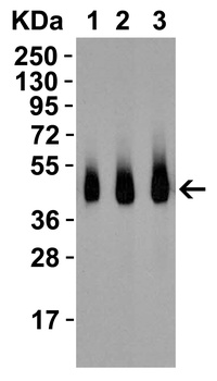
Western Blot Validation with Recombinant Protein. Loading: 30 ng of human RSPO1 recombinant protein per lane. Antibodies: RSPO1, orb1239947 (Lane 1: 0.5 µg/mL, Lane 2: 1 µg/mL, Lane 3: 2 µg/mL), 1h incubation at RT in 5% NFDM/TBST. Secondary: Goat anti-rabbit IgG HRP conjugate at 1:10000 dilution.
Western Blot Validation in Human Spleen. Loading: 15 µg of lysates per lane. Antibodies: RSPO1, orb1239947 (Lane 1: 0.5 µg/mL, Lane 2: 1 µg/mL), 1h incubation at RT in 5% NFDM/TBST. Secondary: Goat anti-rabbit IgG HRP conjugate at 1:10000 dilution.
Western Blot Validation in Human Heart. Loading: 15 µg of lysates per lane. Antibodies: RSPO1, orb1239947 (A: 1 µg/mL, B: 2 µg/mL), 1h incubation at RT in 5% NFDM/TBST. Secondary: Goat anti-rabbit IgG HRP conjugate at 1:10000 dilution.
Immunofluorescence Validation of RSPO1 in Human Spleen. Immunofluorescent analysis of 4% paraformaldehyde-fixed human spleen labeling RSPO1 with orb1239947 at 5 µg/mL, followed by goat anti-rabbit IgG secondary antibody at 1/500 dilution (green) with Phylloidin staining (red) and DAPI staining (blue).
Immunohistochemistry Validation of RSPO1 in Human Heart. Immunohistochemical analysis of paraffin-embedded mouse spleen tissue using anti-RSPO1 antibody (orb1239947) at 2.5 µg/ml. Tissue was fixed with formaldehyde and blocked with 10% serum for 1 h at RT, antigen retrieval was by heat mediation with a citrate buffer (pH6). Samples were incubated with primary antibody overnight at 4C. A goat anti-rabbit IgG H&L (HRP) at 1/250 was used as secondary. Counter stained with Hematoxylin.
Immunohistochemistry Validation of RSPO1 in Human Spleen Tissue. Immunohistochemical analysis of paraffin-embedded human spleen tissue using anti-RSPO1 antibody (orb1239947) at 5 µg/ml. Tissue was fixed with formaldehyde and blocked with 10% serum for 1 h at RT, antigen retrieval was by heat mediation with a citrate buffer (pH6). Samples were incubated with primary antibody overnight at 4C. A goat anti-rabbit IgG H&L (HRP) at 1/250 was used as secondary. Counter stained with Hematoxylin.
Immunohistochemistry Validation of RSPO1 in Human Spleen Tissue. Immunohistochemical analysis of paraffin-embedded human spleen tissue using anti-RSPO1 antibody (orb1239947) at 2.5 µg/ml. Tissue was fixed with formaldehyde and blocked with 10% serum for 1 h at RT, antigen retrieval was by heat mediation with a citrate buffer (pH6). Samples were incubated with primary antibody overnight at 4C. A goat anti-rabbit IgG H&L (HRP) at 1/250 was used as secondary. Counter stained with Hematoxylin.
* Mehrwertsteuer und Versandkosten nicht enthalten. Irrtümer und Preisänderungen vorbehalten