Anti-IL-23 antibody (orb1239493) was raised against a peptide corresponding to 17 amino acids near the amino terminus of human IL-23. The immunogen is located within amino acids 40 - 90 of IL-23.
IL-23 Antibody is supplied in PBS containing 0.02% sodium azide.
Formulierung:
Liquid
Target-Kategorie:
IL23A
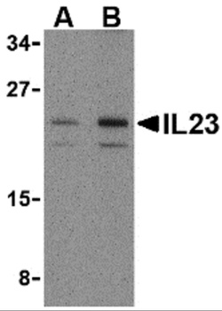
Western Blot Validation in Mouse Pancreas Tissue Lysate. Loading: 15 µg of lysates per lane. Antibodies: IL-23 orb1239493 (A: 1 µg/mL, B: 2 µg/mL), 1h incubation at RT in 5% NFDM/TBST. Secondary: Goat anti-rabbit IgG HRP conjugate at 1:10000 dilution.
Western Blot Validation with Recombinant Protein. Loading: 30 ng of human IL-23 recombinant protein per lane. Antibodies: IL-23 orb1239493 (1: 0.25 µg/mL, 2: 0.5 µg/mL, 3: 1 µg/mL and 4: 2 µg/mL), 1h incubation at RT in 5% NFDM/TBST. Secondary: Goat anti-rabbit IgG HRP conjugate at 1:10000 dilution.
Western Blot Validation in Mouse Tissue Lysate. Loading: 15 µg of lysates per lane. Antibodies: IL-23 orb1239493 (1 µg/mL), 1h incubation at RT in 5% NFDM/TBST. Secondary: Goat anti-rabbit IgG HRP conjugate at 1:10000 dilution.
Immunofluorescence Validation of IL-23 in Mouse Pancreas Tissue. Immunofluorescent analysis of 4% paraformaldehyde-fixed mouse pancreas tissue labeling IL-23 with orb1239493 at 20 µg/mL, followed by goat anti-rabbit IgG secondary antibody at 1/500 dilution (red).
Immunohistochemistry Validation of IL-23 in Mouse Pancreas Tissue. Immunohistochemical analysis of paraffin-embedded mouse pancreas tissue using anti-IL-23 antibody (orb1239493) at 2 µg/ml. Tissue was fixed with formaldehyde and blocked with 10% serum for 1 h at RT, antigen retrieval was by heat mediation with a citrate buffer (pH6). Samples were incubated with primary antibody overnight at 4C. A goat anti-rabbit IgG H&L (HRP) at 1/250 was used as secondary. Counter stained with Hematoxylin.
Induced Expression Validation of IL-23 in Mouse Keratinocytes of Epidermis (Yoon et al., 2016). The expression of IL-23 protein induced by tape stripping was detected by immunohistochemical analysis of mouses skin with anti-IL-23 antibodies. Skin sections from the mouse exhibited epidermis staining positive for IL-23 expression 6 hr after tape stripping. Staining with an isotype-matched irrelevant antibody (Ctrl IgG) was used as a negative control.
* Mehrwertsteuer und Versandkosten nicht enthalten. Irrtümer und Preisänderungen vorbehalten