Anti-BAFF antibody (orb1239254) was raised against a peptide corresponding to 16 amino acids near the carboxy terminus of human BAFF. The immunogen is located within the last 50 amino acids of BAFF.
BAFF Antibody is supplied in PBS containing 0.02% sodium azide.
Formulierung:
Liquid
Target-Kategorie:
TNFSF13B
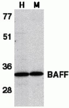
Western Blot Validation in Human HL60 Cell Lysate (H) and Mouse Spleen Lysate (M). Loading: 15 µg of lysates per lane. Antibodies: BAFF orb1239254 (1 µg/mL), 1h incubation at RT in 5% NFDM/TBST. Secondary: Goat anti-rabbit IgG HRP conjugate at 1:10000 dilution.
Western Blot Validation in Human, Mouse and Rat Cell Lines. Loading: 15 µg of lysates per lane. Antibodies: BAFF orb1239254 (1 µg/mL), 1h incubation at RT in 5% NFDM/TBST. Secondary: Goat anti-rabbit IgG HRP conjugate at 1:10000 dilution.
Western Blot Validation with Recombinant Protein. Loading: 30 ng of human BAFF recombinant protein per lane. Antibodies: BAFF orb1239254 (Lane 1: 0.25 µg/mL, Lane 2: 0.5 µg/mL and Lane 3: 1 µg/mL), 1h incubation at RT in 5% NFDM/TBST. Secondary: Goat anti-rabbit IgG HRP conjugate at 1:10000 dilution. Observed at around 18kD.
Immunocytochemistry Validation of BAFF in HL60 Cells. Immunocytochemical analysis of HL60 cells using anti-BAFF antibody (orb1239254) at 1 µg/ml. Cells was fixed with formaldehyde and blocked with 10% serum for 1 h at RT, antigen retrieval was by heat mediation with a citrate buffer (pH6). Samples were incubated with primary antibody overnight at 4C. A goat anti-rabbit IgG H&L (HRP) at 1/250 was used as secondary. Counter stained with Hematoxylin.
Immunofluorescence Validation of BAFF in Human Spleen Tissue. Immunofluorescent analysis of 4% paraformaldehyde-fixed human spleen tissue labeling BAFF with orb1239254 at 20 µg/mL, followed by goat anti-rabbit IgG secondary antibody at 1/500 dilution (green) and DAPI staining (blue).
Regulated Expression Validation of BAFF in Myeloma Patients (Tai et al., 2006). Immunoblot analysis was performed to monitor protein expression of BAFF with anti-BAFF antibodies in multiple myeloma cells with or without BMSCs. BAFF expression in cocultures at 8hr or 24hr was up-regulated by ~3.5-fold relative to BMSCs alone.
Immunohistochemistry Validation of BAFF in Thyroid of Patients with Graves Diseases (Campi et al., 2015). BAFF expression detected by anti-BAFF antibodies (orb1239254) was remarkably increased in thyrocytes from multinodular goiter (C) compared with either Hashimotos thyroiditis (E) or Graves disease (G) while no staining was found in normal thyroid tissue (A).
Immunohistochemistry Validation of BAFF in Murine Cardiac Transplants at Rejection (Ye et al., 2004). BAFF expression detected by anti-BAFF antibodies (orb1239254) was upregulated in intragraft leukocytes due to rejection at 7 days after heart transplant.
* Mehrwertsteuer und Versandkosten nicht enthalten. Irrtümer und Preisänderungen vorbehalten