Boster Bio Anti-Bim BCL2L11 Rabbit Monoclonal Antibody catalog M01552. Tested in WB, IHC, ICC/IF, IP, Flow Cytometry applications. This antibody reacts with Human, Mouse, Rat.
Rabbit IgG in stabilizing components, phosphate buffered saline, pH 7.4, 150mM NaCl, 0.02% sodium azide and 50% glycerol. *This antibody is supplied in a stabilized formulation. Compatibility with conjugation reactions depends on the chemistry of the con
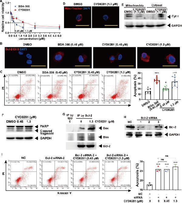
CYD0281 promotes HUVECs cell apoptosis by the exposure of the Bcl-2 BH3 domain. A Cell viability of HUVECs at 48h. The exposure of the BH3 domain of Bcl-2 detected by immunofluorescence (IF) staining using anti-Bcl-2/BH3 domain antibody ( B ), and flow cytometry analysis of cell apoptosis ( C ) in HUVECs treated with BDA-366 and CYD0281 at the IC 50 concentration. HUVECs were treated with CYD0281 at the IC 50 concentration for 48h, mitochondrial dysfunction was analyzed by Mito-Tracker Red CMXRos staining ( D ), Cyt c release ( E ) and PARP cleavage ( F ) were analyzed by western blotting assay, and Bcl-2 associated Bax or Bim was analyzed by co-IP using Bcl-2 antibody ( G ). H The expression of Bcl-2 in HUVECs was inhibited using siRNAs (1, 2, and 3) and analyzed by western blotting assay. I Flow cytometry analysis of cell apoptosis in Bcl-2-silenced HUVECs that were treated with CYD0281 ( n =3). Significant effect compared to the DMSO group: * P <0.05 and *** P <0.001. ns: no significant differences. Scale bars: 50µm Index in PubMed under a CC BY license. PMID: 37237269
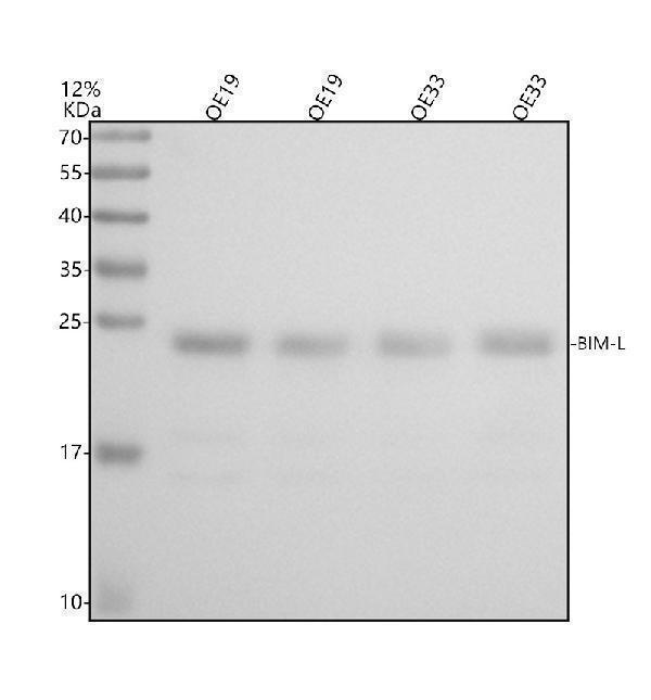
Western blot analysis of Bim using anti-Bim antibody (M01552).Electrophoresis was performed on a 12% SDS-PAGE gel at 80V (Stacking gel) / 120V (Resolving gel) for 2 hours. The sample well of each lane was loaded with 30 µg of sample under reducing conditions.Lane 1: huamn OE19 whole cell lysates,Lane 2: human OE19 whole cell lysates,Lane 3: human OE33 whole cell lysates,Lane 4: human OE33 whole cell lysates.After electrophoresis, proteins were transferred to a nitrocellulose membrane at 150 mA for 50-90 minutes. Blocked the membrane with 5% non-fat milk/TBS for 1.5 hour at RT. The membrane was incubated with rabbit anti-Bim antigen affinity purified monoclonal antibody (M01552) at 0.5 µg/mL overnight at 4C, then washed with TBS-0.1%Tween-20 3 times with 5 minutes each and probed with a goat anti-rabbit IgG-HRP secondary antibody (Catalog BA1054) at a dilution of 1:5000 for 1.5 hour at RT. The signal is developed using an ECL Plus Western Blotting Substrate (Catalog AR1196-200) with Tanon 5200 system. A specific band was detected for Bim-L at approximately ~22 kDa. The expected band size for Bim is at ~22 kDa.
Immunofluorescent analysis using the Antibody at 1:150 dilution.
Immunofluorescent analysis of Raji cells, using Bim Antibody.
Immunohistochemical analysis of paraffin-embedded human cervix cancer, using Bim Antibody.
Immunohistochemical analysis of paraffin-embedded Human prostate cancer, using the Antibody at 1:50 dilution.
Immunohistochemical analysis of paraffin-embedded Human esophageal carcinoma, using the Antibody at 1:50 dilution.
Western blot analysis of Bim expression in A431 cell lysate.
All lanes use the Antibody at 1:1W dilution for 1 hour at room temperature.
* Mehrwertsteuer und Versandkosten nicht enthalten. Irrtümer und Preisänderungen vorbehalten