Boster Bio Anti-LC3B MAP1LC3B Rabbit Monoclonal Antibody catalog M01524. Tested in WB, ICC/IF, IP applications. This antibody reacts with Human, Mouse, Rat.
Rabbit IgG in stabilizing components, phosphate buffered saline, pH 7.4, 150mM NaCl, 0.02% sodium azide and 50% glycerol. *This antibody is supplied in a stabilized formulation. Compatibility with conjugation reactions depends on the chemistry of the con
Reinheit:
Affinity-chromatography
Formulierung:
Liquid
Target-Kategorie:
Microtubule-associated protein 1 light chain 3 beta
Application Verdünnung:
WB 1:500-2000IHC 1:50-200ICC/IF 1:50-200IP 1:50
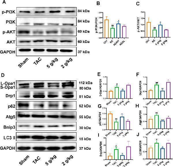
AEA improved CHF via PI3K/AKT/Bnip3 axis. (A) Representative images of PI3K/AKT axis. (B, C) The phosphorylation level of PI3K and AKT. (D) Representative images of Opa1, Drp1, Bnip3, p62, Atg5 and LC3II. (E-J) The expression level of Opa1, Drp1, Bnip3, p62, Atg5 and LC3II. (n = 3).Index in PubMed under a CC BY license. PMID: 40206063
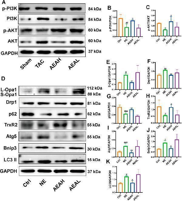
AEA improved NE-induced injuries via PI3K/AKT/Bnip3 axis. (A) Representative images of PI3K/AKT axis in H9c2 cells. (B, C) The phosphorylation level of PI3K and AKT. (D) Representative i
Immunofluorescent analysis using the Antibody at 1:150 dilution.
Immunofluorescent analysis of Hela cells treated with choroquine, using LC3B Antibody.
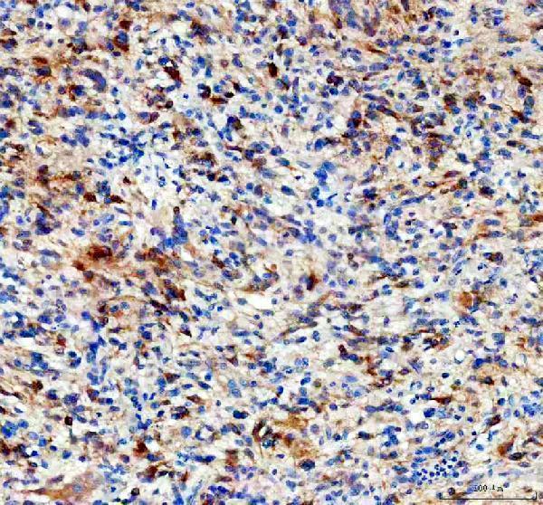
IHC analysis of LC3B using anti-LC3B antibody (M01524). LC3B was detected in a paraffin-embedded section of human glioma tissue. Heat mediated antigen retrieval was performed in EDTA buffer (pH 8.0, epitope retrieval solution). The tissue section was blocked with 10% goat serum. The tissue section was then incubated with 1:50 rabbit anti-LC3B Antibody (M01524) overnight at 4C. Peroxidase Conjugated Goat Anti-rabbit IgG was used as secondary antibody and incubated for 30 minutes at 37C. The tissue section was developed using HRP Conjugated Rabbit IgG Super Vision Assay Kit (Catalog SV0002) with DAB as the chromogen.
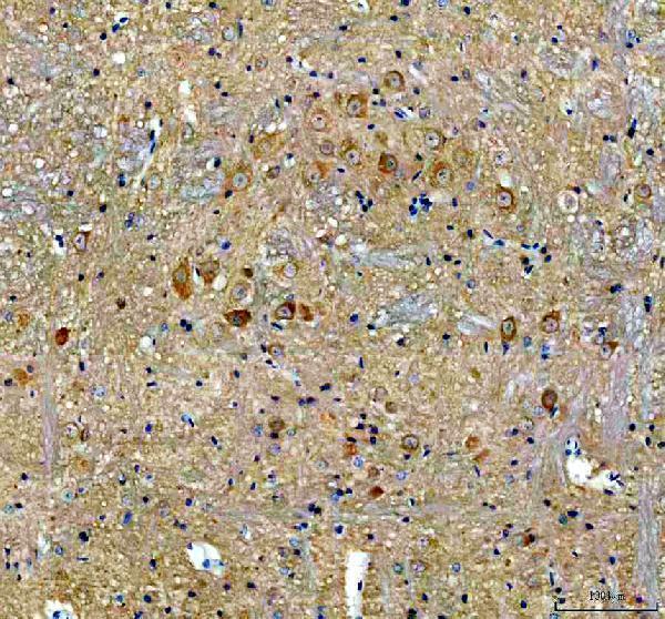
IHC analysis of LC3B using anti-LC3B antibody (M01524). LC3B was detected in a paraffin-embedded section of mouse brain tissue. Heat mediated antigen retrieval was performed in EDTA buffer (pH 8.0, epitope retrieval solution). The tissue section was blocked with 10% goat serum. The tissue section was then incubated with 1:50 rabbit anti-LC3B Antibody (M01524) overnight at 4C. Peroxidase Conjugated Goat Anti-rabbit IgG was used as secondary antibody and incubated for 30 minutes at 37C. The tissue section was developed using HRP Conjugated Rabbit IgG Super Vision Assay Kit (Catalog SV0002) with DAB as the chromogen.
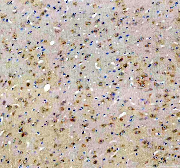
IHC analysis of LC3B using anti-LC3B antibody (M01524). LC3B was detected in a paraffin-embedded section of rat brain tissue. Heat mediated antigen retrieval was performed in EDTA buffer (pH 8.0, epitope retrieval solution). The tissue section was blocked with 10% goat serum. The tissue section was then incubated with 1:50 rabbit anti-LC3B Antibody (M01524) overnight at 4C. Peroxidase Conjugated Goat Anti-rabbit IgG was used as secondary antibody and incubated for 30 minutes at 37C. The tissue section was developed using HRP Conjugated Rabbit IgG Super Vision Assay Kit (Catalog SV0002) with DAB as the chromogen.
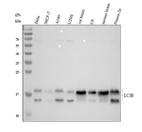
Western blot analysis of LC3B using anti-LC3B antibody (M01524). Electrophoresis was performed on a 12% SDS-PAGE gel at 80V (Stacking gel) / 120V (Resolving gel) for 2 hours. The sample well of each lane was loaded with 30 ug of sample under reducing conditions. Lane 1: human Hela whole cell lysates,Lane 2: human MCF-7 whole cell lysates,Lane 3: human A549 whole cell lysates,Lane 4: human U2OS whole cell lysates,Lane 5: rat brain tissue lysates,Lane 6: rat C6 whole cell lysates,Lane 7: mouse brain tissue lysates,Lane 8: mouse Neurao-2a whole cell lysates.After electrophoresis, proteins were transferred to a nitrocellulose membrane at 150 mA for 50-90 minutes. Blocked the membrane with 5% non-fat milk/TBS for 1.5 hour at RT. The membrane was incubated with rabbit anti-LC3B antigen affinity purified monoclonal antibody (Catalog M01524) at 1:500 overnight at 4C, then washed with TBS-0.1%Tween 3 times with 5 minutes each and probed with a goat anti-rabbit IgG-HRP secondary antibody at a dilution of 1:500 for 1.5 hour at RT. The signal is developed using an ECL Plus Western Blotting Substrate (Catalog AR1196-200) with Tanon 5200 system. A specific band was detected for LC3B at approximately 15, 18 kDa. The expected band size for LC3B is at 15 kDa.
* Mehrwertsteuer und Versandkosten nicht enthalten. Irrtümer und Preisänderungen vorbehalten