Boster Bio Anti-NGFR/P75 Rabbit Monoclonal Antibody catalog M01187-1. Tested in WB, IHC, ICC/IF, IP applications. This antibody reacts with Human, Mouse, Rat.
Rabbit IgG in stabilizing components, phosphate buffered saline, pH 7.4, 150mM NaCl, 0.02% sodium azide and 50% glycerol. *This antibody is supplied in a stabilized formulation. Compatibility with conjugation reactions depends on the chemistry of the con
Reinheit:
Affinity-chromatography
Formulierung:
Liquid
Target-Kategorie:
Tumor necrosis factor receptor superfamily member 16
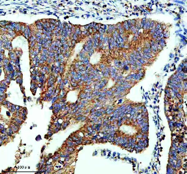
IHC analysis of NGFR using anti-NGFR antibody (M01187-1). NGFR was detected in a paraffin-embedded section of human colon cancer tissue. Heat mediated antigen retrieval was performed in EDTA buffer (pH 8.0, epitope retrieval solution). The tissue section was blocked with 10% goat serum. The tissue section was then incubated with 1:50 rabbit anti-NGFR Antibody (M01187-1) overnight at 4C. Peroxidase Conjugated Goat Anti-rabbit IgG was used as secondary antibody and incubated for 30 minutes at 37C. The tissue section was developed using HRP Conjugated Rabbit IgG Super Vision Assay Kit (Catalog SV0002) with DAB as the chromogen.
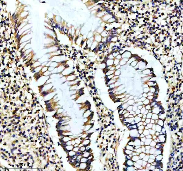
IHC analysis of NGFR using anti-NGFR antibody (M01187-1). NGFR was detected in a paraffin-embedded section of human colon tissue. Heat mediated antigen retrieval was performed in EDTA buffer (pH 8.0, epitope retrieval solution). The tissue section was blocked with 10% goat serum. The tissue section was then incubated with 1:50 rabbit anti-NGFR Antibody (M01187-1) overnight at 4C. Peroxidase Conjugated Goat Anti-rabbit IgG was used as secondary antibody and incubated for 30 minutes at 37C. The tissue section was developed using HRP Conjugated Rabbit IgG Super Vision Assay Kit (Catalog SV0002) with DAB as the chromogen.
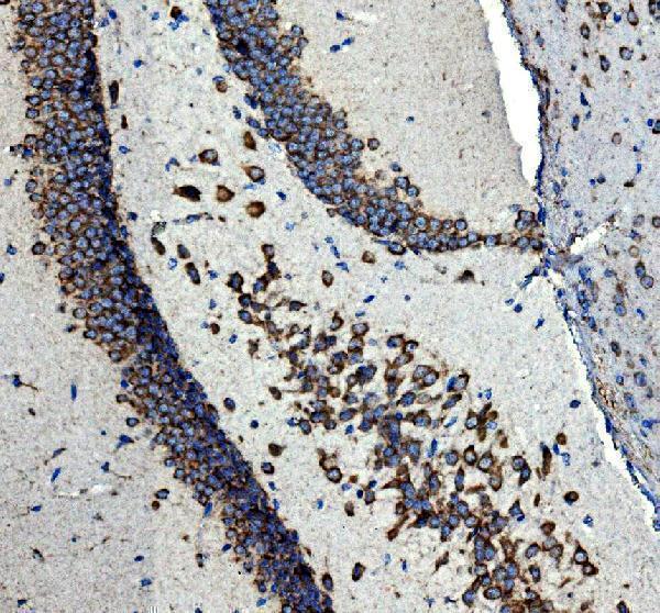
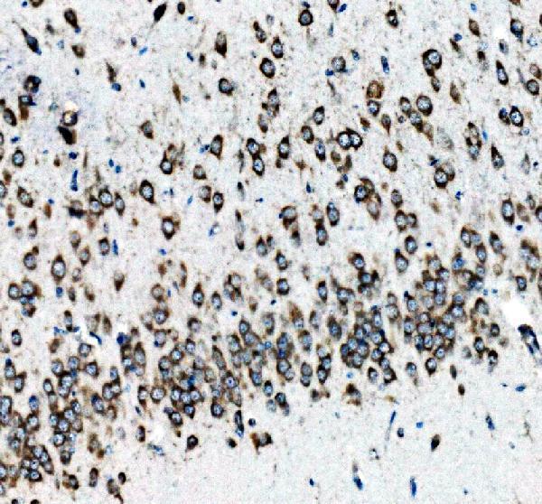
IHC analysis of NGFR using anti-NGFR antibody (M01187-1). NGFR was detected in a paraffin-embedded section of mouse brain tissue. Heat mediated antigen retrieval was performed in EDTA buffer (pH 8.0, epitope retrieval solution). The tissue section was blocked with 10% goat serum. The tissue section was then incubated with 1:50 rabbit anti-NGFR Antibody (M01187-1) overnight at 4C. Peroxidase Conjugated Goat Anti-rabbit IgG was used as secondary antibody and incubated for 30 minutes at 37C. The tissue section was developed using HRP Conjugated Rabbit IgG Super Vision Assay Kit (Catalog SV0002) with DAB as the chromogen.
IHC analysis of NGFR using anti-NGFR antibody (M01187-1). NGFR was detected in a paraffin-embedded section of mouse brain tissue. Heat mediated antigen retrieval was performed in EDTA buffer (pH 8.0, epitope retrieval solution). The tissue section was blocked with 10% goat serum. The tissue section was then incubated with 1:50 rabbit anti-NGFR Antibody (M01187-1) overnight at 4C. Peroxidase Conjugated Goat Anti-rabbit IgG was used as secondary antibody and incubated for 30 minutes at 37C. The tissue section was developed using HRP Conjugated Rabbit IgG Super Vision Assay Kit (Catalog SV0002) with DAB as the chromogen.
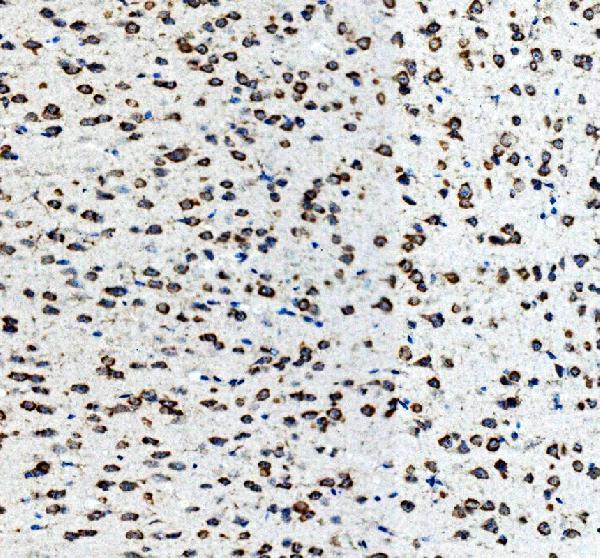
IHC analysis of NGFR using anti-NGFR antibody (M01187-1). NGFR was detected in a paraffin-embedded section of rat brain tissue. Heat mediated antigen retrieval was performed in EDTA buffer (pH 8.0, epitope retrieval solution). The tissue section was blocked with 10% goat serum. The tissue section was then incubated with 1:50 rabbit anti-NGFR Antibody (M01187-1) overnight at 4C. Peroxidase Conjugated Goat Anti-rabbit IgG was used as secondary antibody and incubated for 30 minutes at 37C. The tissue section was developed using HRP Conjugated Rabbit IgG Super Vision Assay Kit (Catalog SV0002) with DAB as the chromogen.
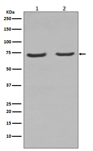
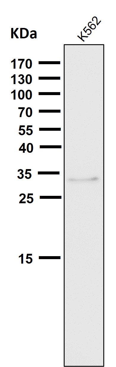
Western blot analysis of NGFR expression in (1) C6 cell lysate, (2) PC-12 cell lysate.
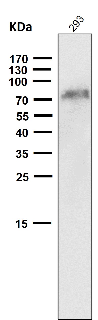
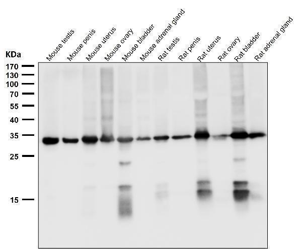
All lanes use the Antibody at 1:1W dilution for 1 hour at room temperature.
All lanes use the Antibody at 1:1W dilution for 1 hour at room temperature.
All lanes use the Antibody at 1:1W dilution for 1 hour at room temperature.
* Mehrwertsteuer und Versandkosten nicht enthalten. Irrtümer und Preisänderungen vorbehalten